5J19 | pdb_00005j19

phospho-Pon binding-induced Plk1 dimerization


Experimental Data Snapshot

  • Method: X-RAY DIFFRACTION
  • Resolution: 2.00 Å
  • R-Value Free: 
    0.236 (Depositor), 0.236 (DCC) 
  • R-Value Work: 
    0.213 (Depositor), 0.213 (DCC) 
  • R-Value Observed: 
    0.214 (Depositor) 

Starting Model: experimental
View more details

wwPDB Validation 3D Report Full Report

Validation slider image for 5J19

This is version 1.3 of the entry. See complete history

Literature

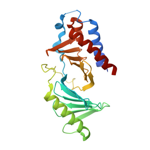
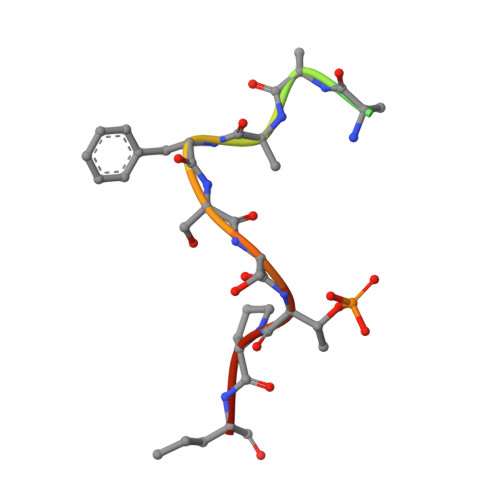
Phospho-Pon Binding-Mediated Fine-Tuning of Plk1 Activity

Zhu, K.Shan, Z.Zhang, L.Wen, W.

(2016) Structure 24: 1110-1119

  • DOI: https://doi.org/10.1016/j.str.2016.04.012
  • Primary Citation Related Structures: 
    5J19

  • PubMed Abstract: 

    In Drosophila neuroblasts (NBs), the asymmetrical localization and segregation of the cell-fate determinant Numb are regulated by its adaptor Partner of Numb (Pon) and the cell-cycle kinase Polo. Polo phosphorylates the Pon localization domain, thus leading to its basal distribution together with Numb, albeit through an unclear mechanism. Here, we find that Cdk1 phosphorylates Pon at Thr63, thus creating a docking site for the Polo-box domain (PBD) of Polo-like kinase 1 (Plk1). The crystal structure of the Plk1 PBD/phospho-Pon complex reveals that two phospho-Pon bound PBDs associate to form a dimer of dimers. We provide evidence that phospho-Pon binding-induced PBD dimerization relieves the autoinhibition of Plk1. Moreover, we demonstrate that the priming Cdk1 phosphorylation of Pon is important for sequential Plk1 phosphorylation. Our results not only provide structural insight into how phosphoprotein binding activates Plk1 but also suggest that binding to different phosphoproteins might mediate the fine-tuning of Plk1 activity.


  • Organizational Affiliation
    • Department of Neurosurgery, Huashan Hospital, Institutes of Biomedical Sciences, Fudan University, Shanghai 200040, China.

Macromolecule Content 

  • Total Structure Weight: 57.54 kDa 
  • Atom Count: 3,705 
  • Modeled Residue Count: 468 
  • Deposited Residue Count: 498 
  • Unique protein chains: 2

Macromolecules

Find similar proteins by:|  3D Structure
Entity ID: 1
MoleculeChains  Sequence LengthOrganismDetailsImage
Serine/threonine-protein kinase PLK1
A, B
234Homo sapiensMutation(s): 0 
Gene Names: PLK1PLK
EC: 2.7.11.21
UniProt & NIH Common Fund Data Resources
Find proteins for P53350 (Homo sapiens)
Explore P53350 
Go to UniProtKB:  P53350
PHAROS:  P53350
GTEx:  ENSG00000166851 
Entity Groups
Sequence Clusters30% Identity50% Identity70% Identity90% Identity95% Identity100% Identity
UniProt GroupP53350
Sequence Annotations
Expand
Reference Sequence
Find similar proteins by:  Sequence   |   3D Structure  
Entity ID: 2
MoleculeChains  Sequence LengthOrganismDetailsImage
Phosphorylated peptide from Partner of Numb
C, D
15Drosophila melanogasterMutation(s): 0 
UniProt
Find proteins for O96561 (Drosophila melanogaster)
Explore O96561 
Go to UniProtKB:  O96561
Entity Groups
Sequence Clusters30% Identity50% Identity70% Identity90% Identity95% Identity100% Identity
UniProt GroupO96561
Sequence Annotations
Expand
Reference Sequence

Experimental Data & Validation

Experimental Data

  • Method: X-RAY DIFFRACTION
  • Resolution: 2.00 Å
  • R-Value Free:  0.236 (Depositor), 0.236 (DCC) 
  • R-Value Work:  0.213 (Depositor), 0.213 (DCC) 
  • R-Value Observed: 0.214 (Depositor) 
Space Group: C 2 2 21
Unit Cell:
Length ( Å )Angle ( ˚ )
a = 84.88α = 90
b = 91.997β = 90
c = 159.222γ = 90
Software Package:
Software NamePurpose
HKL-2000data collection
HKL-2000data scaling
PHENIXrefinement
PDB_EXTRACTdata extraction
HKL-2000data reduction
PHASERphasing

Structure Validation

View Full Validation Report


Entry History 

Deposition Data

Revision History  (Full details and data files)

  • Version 1.0: 2016-06-15
    Type: Initial release
  • Version 1.1: 2016-07-20
    Changes: Database references
  • Version 1.2: 2023-11-08
    Changes: Data collection, Database references, Derived calculations, Refinement description
  • Version 1.3: 2024-10-09
    Changes: Structure summary