5J0E

Crystal structures of Pribnow box consensus promoter sequence (P32)


Experimental Data Snapshot

  • Method: X-RAY DIFFRACTION
  • Resolution: 2.81 Å
  • R-Value Free: 0.248 
  • R-Value Work: 0.226 
  • R-Value Observed: 0.227 

wwPDB Validation   3D Report Full Report


This is version 1.2 of the entry. See complete history


Literature

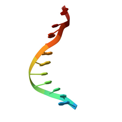
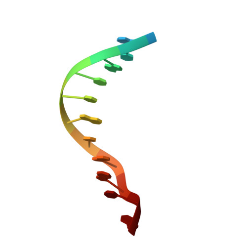
Structure elucidation of the Pribnow box consensus promoter sequence by racemic DNA crystallography.

Mandal, P.K.Collie, G.W.Srivastava, S.C.Kauffmann, B.Huc, I.

(2016) Nucleic Acids Res 44: 5936-5943

  • DOI: https://doi.org/10.1093/nar/gkw367
  • Primary Citation of Related Structures:  
    5ET9, 5EWB, 5EYQ, 5EZF, 5F26, 5J0E

  • PubMed Abstract: 

    It has previously been shown that the use of racemic mixtures of naturally chiral macromolecules such as protein and DNA can significantly aid the crystallogenesis process, thereby addressing one of the major bottlenecks to structure determination by X-ray crystallographic methods-that of crystal growth. Although previous studies have provided convincing evidence of the applicability of the racemic crystallization technique to DNA through the study of well-characterized DNA structures, we sought to apply this method to a historically challenging DNA sequence. For this purpose we chose a non-self-complementary DNA duplex containing the biologically-relevant Pribnow box consensus sequence 'TATAAT'. Four racemic crystal structures of this previously un-crystallizable DNA target are reported (with resolutions in the range of 1.65-2.3 Å), with further crystallographic studies and structural analysis providing insight into the racemic crystallization process as well as structural details of this highly pertinent DNA sequence.


  • Organizational Affiliation

    Université de Bordeaux, CBMN (UMR5248), Pessac 33600, France CNRS, CBMN (UMR5248), Pessac 33600, France.


Macromolecules

Find similar nucleic acids by:  Sequence   |   3D Structure  

Entity ID: 1
MoleculeChains LengthOrganismImage
Pribnow box consensus promoter sequence
A, C, E
12synthetic construct
Sequence Annotations
Expand
  • Reference Sequence

Find similar nucleic acids by:  Sequence   |   3D Structure  

Entity ID: 2
MoleculeChains LengthOrganismImage
Complementary sequence
B, D, F
12synthetic construct
Sequence Annotations
Expand
  • Reference Sequence
Experimental Data & Validation

Experimental Data

  • Method: X-RAY DIFFRACTION
  • Resolution: 2.81 Å
  • R-Value Free: 0.248 
  • R-Value Work: 0.226 
  • R-Value Observed: 0.227 
  • Space Group: P 32
Unit Cell:
Length ( Å )Angle ( ˚ )
a = 46.906α = 90
b = 46.906β = 90
c = 99.044γ = 120
Software Package:
Software NamePurpose
REFMACrefinement
XDSdata reduction
XDSdata scaling
PHASERphasing

Structure Validation

View Full Validation Report


Entry History & Funding Information

Deposition Data


Funding OrganizationLocationGrant Number
European Research CouncilFranceERC-2012-AdG-320892

Revision History  (Full details and data files)

  • Version 1.0: 2016-05-18
    Type: Initial release
  • Version 1.1: 2016-07-20
    Changes: Database references
  • Version 1.2: 2024-01-10
    Changes: Data collection, Database references, Derived calculations, Refinement description