5IY0

PfMCM N-terminal domain double hexamer


Experimental Data Snapshot

  • Method: X-RAY DIFFRACTION
  • Resolution: 3.00 Å
  • R-Value Free: 0.223 
  • R-Value Work: 0.205 
  • R-Value Observed: 0.205 

wwPDB Validation   3D Report Full Report


This is version 1.4 of the entry. See complete history


Literature

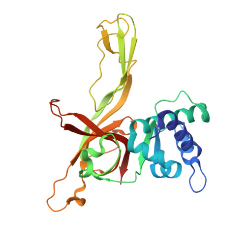
Structure of a double hexamer of the Pyrococcus furiosus minichromosome maintenance protein N-terminal domain.

Meagher, M.Enemark, E.J.

(2016) Acta Crystallogr F Struct Biol Commun 72: 545-551

  • DOI: https://doi.org/10.1107/S2053230X1600858X
  • Primary Citation of Related Structures:  
    5IY0

  • PubMed Abstract: 

    The crystal structure of the N-terminal domain of the Pyrococcus furiosus minichromosome maintenance (MCM) protein as a double hexamer is described. The MCM complex is a ring-shaped helicase that unwinds DNA at the replication fork of eukaryotes and archaea. Prior to replication initiation, the MCM complex assembles as an inactive double hexamer at specific sites of DNA. The presented structure is highly consistent with previous MCM double-hexamer structures and shows two MCM hexamers with a head-to-head interaction mediated by the N-terminal domain. Minor differences include a diminished head-to-head interaction and a slightly reduced inter-hexamer rotation.


  • Organizational Affiliation

    Department of Structural Biology, St Jude Children's Research Hospital, 262 Danny Thomas Place, Memphis, TN 38105, USA.


Macromolecules
Find similar proteins by:  (by identity cutoff)  |  3D Structure
Entity ID: 1
MoleculeChains Sequence LengthOrganismDetailsImage
Cell division control protein 21
A, B, C, D, E
A, B, C, D, E, F
257Pyrococcus furiosus DSM 3638Mutation(s): 0 
Gene Names: PF0482
UniProt
Find proteins for Q8U3I4 (Pyrococcus furiosus (strain ATCC 43587 / DSM 3638 / JCM 8422 / Vc1))
Explore Q8U3I4 
Go to UniProtKB:  Q8U3I4
Entity Groups  
Sequence Clusters30% Identity50% Identity70% Identity90% Identity95% Identity100% Identity
UniProt GroupQ8U3I4
Sequence Annotations
Expand
  • Reference Sequence
Experimental Data & Validation

Experimental Data

  • Method: X-RAY DIFFRACTION
  • Resolution: 3.00 Å
  • R-Value Free: 0.223 
  • R-Value Work: 0.205 
  • R-Value Observed: 0.205 
  • Space Group: C 1 2 1
Unit Cell:
Length ( Å )Angle ( ˚ )
a = 211.427α = 90
b = 122.61β = 92.35
c = 111.892γ = 90
Software Package:
Software NamePurpose
REFMACrefinement
HKL-2000data reduction
HKL-2000data scaling
PHASERphasing

Structure Validation

View Full Validation Report


Entry History & Funding Information

Deposition Data


Funding OrganizationLocationGrant Number
National Institutes of Health/National Institute of General Medical Sciences (NIH/NIGMS)United StatesR01-GM098771

Revision History  (Full details and data files)

  • Version 1.0: 2016-07-13
    Type: Initial release
  • Version 1.1: 2016-07-20
    Changes: Database references
  • Version 1.2: 2017-09-27
    Changes: Author supporting evidence, Derived calculations
  • Version 1.3: 2019-12-25
    Changes: Author supporting evidence
  • Version 1.4: 2023-09-27
    Changes: Data collection, Database references, Refinement description