5IX1

Crystal structure of mouse Morc3 ATPase-CW cassette in complex with AMPPNP and H3K4me3 peptide


Experimental Data Snapshot

  • Method: X-RAY DIFFRACTION
  • Resolution: 2.60 Å
  • R-Value Free: 0.230 
  • R-Value Work: 0.218 
  • R-Value Observed: 0.218 

wwPDB Validation   3D Report Full Report


Ligand Structure Quality Assessment 


This is version 1.1 of the entry. See complete history


Literature

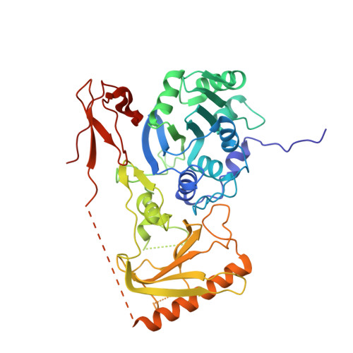
Mouse MORC3 is a GHKL ATPase that localizes to H3K4me3 marked chromatin

Li, S.Yen, L.Pastor, W.A.Johnston, J.B.Du, J.Shew, C.J.Liu, W.Ho, J.Stender, B.Clark, A.T.Burlingame, A.L.Daxinger, L.Patel, D.J.Jacobsen, S.E.

(2016) Proc Natl Acad Sci U S A 113: E5108-E5116

  • DOI: https://doi.org/10.1073/pnas.1609709113
  • Primary Citation of Related Structures:  
    5IX1, 5IX2

  • PubMed Abstract: 

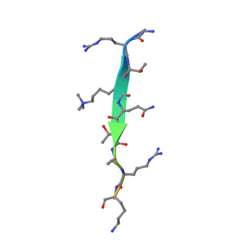
    Microrchidia (MORC) proteins are GHKL (gyrase, heat-shock protein 90, histidine kinase, MutL) ATPases that function in gene regulation in multiple organisms. Animal MORCs also contain CW-type zinc finger domains, which are known to bind to modified histones. We solved the crystal structure of the murine MORC3 ATPase-CW domain bound to the nucleotide analog AMPPNP (phosphoaminophosphonic acid-adenylate ester) and in complex with a trimethylated histone H3 lysine 4 (H3K4) peptide (H3K4me3). We observed that the MORC3 N-terminal ATPase domain forms a dimer when bound to AMPPNP. We used native mass spectrometry to show that dimerization is ATP-dependent, and that dimer formation is enhanced in the presence of nonhydrolyzable ATP analogs. The CW domain uses an aromatic cage to bind trimethylated Lys4 and forms extensive hydrogen bonds with the H3 tail. We found that MORC3 localizes to promoters marked by H3K4me3 throughout the genome, consistent with its binding to H3K4me3 in vitro. Our work sheds light on aspects of the molecular dynamics and function of MORC3.


  • Organizational Affiliation

    Structural Biology Program, Memorial Sloan Kettering Cancer Center, New York, NY 10065;


Macromolecules
Find similar proteins by:  (by identity cutoff)  |  3D Structure
Entity ID: 1
MoleculeChains Sequence LengthOrganismDetailsImage
MORC family CW-type zinc finger protein 3
A, B
451Mus musculusMutation(s): 0 
Gene Names: Morc3
UniProt & NIH Common Fund Data Resources
Find proteins for F7BJB9 (Mus musculus)
Explore F7BJB9 
Go to UniProtKB:  F7BJB9
IMPC:  MGI:2136841
Entity Groups  
Sequence Clusters30% Identity50% Identity70% Identity90% Identity95% Identity100% Identity
UniProt GroupF7BJB9
Sequence Annotations
Expand
  • Reference Sequence

Find similar proteins by:  Sequence   |   3D Structure  

Entity ID: 2
MoleculeChains Sequence LengthOrganismDetailsImage
Peptide from Histone H3.1C [auth P],
D [auth Q]
15Mus musculusMutation(s): 0 
UniProt
Find proteins for P68433 (Mus musculus)
Explore P68433 
Go to UniProtKB:  P68433
Entity Groups  
Sequence Clusters30% Identity50% Identity70% Identity90% Identity95% Identity100% Identity
UniProt GroupP68433
Sequence Annotations
Expand
  • Reference Sequence
Experimental Data & Validation

Experimental Data

  • Method: X-RAY DIFFRACTION
  • Resolution: 2.60 Å
  • R-Value Free: 0.230 
  • R-Value Work: 0.218 
  • R-Value Observed: 0.218 
  • Space Group: I 2 2 2
Unit Cell:
Length ( Å )Angle ( ˚ )
a = 111.353α = 90
b = 149.079β = 90
c = 173.473γ = 90
Software Package:
Software NamePurpose
PHENIXrefinement
HKL-2000data reduction
HKL-2000data scaling
PHENIXphasing

Structure Validation

View Full Validation Report


Ligand Structure Quality Assessment 


Entry History 

Deposition Data

Revision History  (Full details and data files)

  • Version 1.0: 2016-08-17
    Type: Initial release
  • Version 1.1: 2016-09-14
    Changes: Database references