5IEY | pdb_00005iey

Crystal structure of a CDK inhibitor bound to CDK2


Experimental Data Snapshot

  • Method: X-RAY DIFFRACTION
  • Resolution: 1.66 Å
  • R-Value Free: 
    0.225 (Depositor), 0.250 (DCC) 
  • R-Value Work: 
    0.180 (Depositor) 
  • R-Value Observed: 
    0.182 (Depositor) 

wwPDB Validation 3D Report Full Report

Validation slider image for 5IEY

Ligand Structure Quality Assessment 


This is version 1.4 of the entry. See complete history

Literature

Conformational Adaption May Explain the Slow Dissociation Kinetics of Roniciclib (BAY 1000394), a Type I CDK Inhibitor with Kinetic Selectivity for CDK2 and CDK9.

Ayaz, P.Andres, D.Kwiatkowski, D.A.Kolbe, C.C.Lienau, P.Siemeister, G.Lucking, U.Stegmann, C.M.

(2016) ACS Chem Biol 11: 1710-1719

  • DOI: https://doi.org/10.1021/acschembio.6b00074
  • Primary Citation Related Structures: 
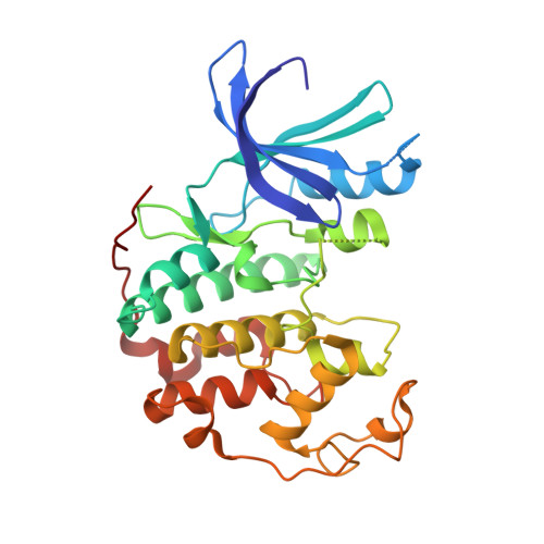
    5IEV, 5IEX, 5IEY, 5IF1

  • PubMed Abstract: 

    Roniciclib (BAY 1000394) is a type I pan-CDK (cyclin-dependent kinase) inhibitor which has revealed potent efficacy in xenograft cancer models. Here, we show that roniciclib displays prolonged residence times on CDK2 and CDK9, whereas residence times on other CDKs are transient, thus giving rise to a kinetic selectivity of roniciclib. Surprisingly, variation of the substituent at the 5-position of the pyrimidine scaffold results in changes of up to 3 orders of magnitude of the drug-target residence time. CDK2 X-ray cocrystal structures have revealed a DFG-loop adaption for the 5-(trifluoromethyl) substituent, while for hydrogen and bromo substituents the DFG loop remains in its characteristic type I inhibitor position. In tumor cells, the prolonged residence times of roniciclib on CDK2 and CDK9 are reflected in a sustained inhibitory effect on retinoblastoma protein (RB) phosphorylation, indicating that the target residence time on CDK2 may contribute to sustained target engagement and antitumor efficacy.


  • Organizational Affiliation
    • Bayer Pharma AG , Drug Discovery, Lead Discovery Berlin, Berlin, Germany.

Macromolecule Content 

  • Total Structure Weight: 34.31 kDa 
  • Atom Count: 2,557 
  • Modeled Residue Count: 286 
  • Deposited Residue Count: 298 
  • Unique protein chains: 1

Macromolecules

Find similar proteins by:|  3D Structure
Entity ID: 1
MoleculeChains  Sequence LengthOrganismDetailsImage
Cyclin-dependent kinase 2298Homo sapiensMutation(s): 0 
Gene Names: CDK2CDKN2
EC: 2.7.11.22
UniProt & NIH Common Fund Data Resources
Find proteins for P24941 (Homo sapiens)
Explore P24941 
Go to UniProtKB:  P24941
PHAROS:  P24941
GTEx:  ENSG00000123374 
Entity Groups
Sequence Clusters30% Identity50% Identity70% Identity90% Identity95% Identity100% Identity
UniProt GroupP24941
Sequence Annotations
Expand
Reference Sequence

Small Molecules

Ligands 1 Unique
IDChains Name / Formula / InChI Key2D Diagram3D Interactions
6AE

Query on 6AE


Download:Ideal Coordinates CCD File
B [auth A]4-[(4-{[(2R,3R)-3-hydroxybutan-2-yl]amino}pyrimidin-2-yl)amino]benzene-1-sulfonamide
C14 H19 N5 O3 S
BOQCFLZSDDGZTL-NXEZZACHSA-N

Experimental Data & Validation

Experimental Data

  • Method: X-RAY DIFFRACTION
  • Resolution: 1.66 Å
  • R-Value Free:  0.225 (Depositor), 0.250 (DCC) 
  • R-Value Work:  0.180 (Depositor) 
  • R-Value Observed: 0.182 (Depositor) 
Space Group: P 21 21 21
Unit Cell:
Length ( Å )Angle ( ˚ )
a = 53.337α = 90
b = 71.983β = 90
c = 72.037γ = 90
Software Package:
Software NamePurpose
REFMACrefinement
XDSdata reduction
XSCALEdata scaling
PHASERphasing

Structure Validation

View Full Validation Report


Ligand Structure Quality Assessment 


Entry History 

Deposition Data

Revision History  (Full details and data files)

  • Version 1.0: 2016-04-27
    Type: Initial release
  • Version 1.1: 2016-05-04
    Changes: Database references
  • Version 1.2: 2016-06-29
    Changes: Database references
  • Version 1.3: 2019-10-16
    Changes: Data collection
  • Version 1.4: 2024-05-08
    Changes: Data collection, Database references