Time-dependent X-ray diffraction studies on urea/hen egg white lysozyme complexes reveal structural changes that indicate onset of denaturation
Raskar, T., Khavnekar, S., Hosur, M.(2016) Sci Rep 6: 32277-32277
- PubMed: 27573790 Search on PubMedSearch on PubMed Central
- DOI: https://doi.org/10.1038/srep32277
- Primary Citation Related Structures:
5I4W, 5I4X, 5I4Y, 5I53, 5I54 - PubMed Abstract:
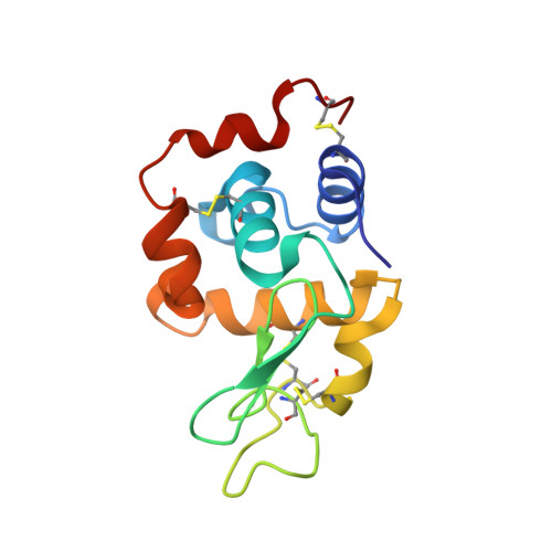
Temporal binding of urea to lysozyme was examined using X-ray diffraction of single crystals of urea/lysozyme complexes prepared by soaking native lysozyme crystals in solutions containing 9 M urea. Four different soak times of 2, 4, 7 and 10 hours were used. The five crystal structures (including the native lysozyme), refined to 1.6 Å resolution, reveal that as the soaking time increased, more and more first-shell water molecules are replaced by urea. The number of hydrogen bonds between urea and the protein is similar to that between protein and water molecules replaced by urea. However, the number of van der Waals contacts to protein from urea is almost double that between the protein and the replaced water. The hydrogen bonding and van der Waals interactions are initially greater with the backbone and later with side chains of charged residues. Urea altered the water-water hydrogen bond network both by replacing water solvating hydrophobic residues and by shortening the first-shell intra-water hydrogen bonds by 0.2 Å. These interaction data suggest that urea uses both 'direct' and 'indirect' mechanisms to unfold lysozyme. Specific structural changes constitute the first steps in lysozyme unfolding by urea.
- Tata Memorial Centre/Advanced Centre for Treatment Research and Education in Cancer, Kharghar, Navi Mumbai, 410210, India.
Organizational Affiliation: