Role of the Tyr270 residue in 2-methyl-3-hydroxypyridine-5-carboxylic acid oxygenase from Mesorhizobium loti
Kobayashi, J., Yoshida, H., Yagi, T., Kamitori, S., Hayashi, H., Mizutani, K., Takahashi, N., Mikami, B.(2017) J Biosci Bioeng 123: 154-162
- PubMed: 27568368 Search on PubMed
- DOI: https://doi.org/10.1016/j.jbiosc.2016.07.022
- Primary Citation Related Structures:
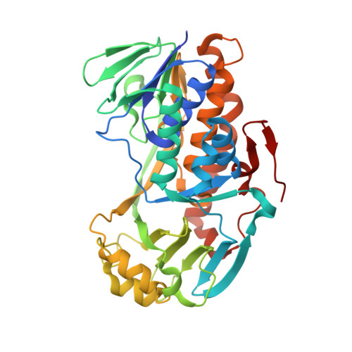
5HXI - PubMed Abstract:
The flavoenzyme 2-Methyl-3-hydroxypyridine-5-carboxylic acid oxygenase (MHPCO) catalyzes the cleavage of the pyridine ring of 2-methyl-3-hydroxypyridine-5-carboxylic acid (MHPC) in the presence of NADH, molecular oxygen, and water. MHPCO also catalyzes the NADH oxidation reaction uncoupled with ring opening in the absence of MHPC (the basal activity). The enzyme shows activity toward not only MHPC but also 5-hydroxynicotinic acid (5HN) and 5-pyridoxic acid (5PA). The reaction rate toward 5PA is extremely low (5% of the activity toward MHPC or 5HN). We determined the crystal structures of MHPCO without substrate and the MHPCO/5HN and MHPCO/5PA complexes, together with a Y270F mutant without substrate and its 5HN complex. The Tyr270 residue was located in the active site and formed hydrogen bonds between the Oη and water molecules to make the active site hydrophilic. Although Tyr270 took a fixed conformation in the structures of the MHPCO and MHPCO/5HN complex, it took two conformations in its 5PA complex, accompanied by two conformations of the bound 5PA. In the wild-type (WT) enzyme, the turnover number of the ring-opening activity was 6800 times that of the basal activity (1300 and 0.19 s -1 , respectively), whereas no such difference was observed in the Y270F (19 and 7.4 s -1 ) or Y270A (0.05 and 0.84 s -1 ) mutants. In the Y270F/5HN complex, the substrate bound ∼1 Å farther away than in the WT enzyme. These results revealed that Tyr270 is essential to maintain the WT conformation, which in turn enhances the coupling of the NADH oxidation with the ring-opening reaction.
- Laboratory of Applied Structural Biology, Division of Applied Life Sciences, Graduate School of Agriculture, Kyoto University, Gokasyo, Uji, Kyoto 611-0011, Japan. Electronic address: jk_virtue_0216@yahoo.co.jp.
Organizational Affiliation: