5HRT | pdb_00005hrt

Crystal structure of mouse autotaxin in complex with a DNA aptamer


Experimental Data Snapshot

  • Method: X-RAY DIFFRACTION
  • Resolution: 2.00 Å
  • R-Value Free: 
    0.202 (Depositor), 0.203 (DCC) 
  • R-Value Work: 
    0.164 (Depositor), 0.166 (DCC) 
  • R-Value Observed: 
    0.165 (Depositor) 

wwPDB Validation 3D Report Full Report

Validation slider image for 5HRT

This is version 2.1 of the entry. See complete history

Literature

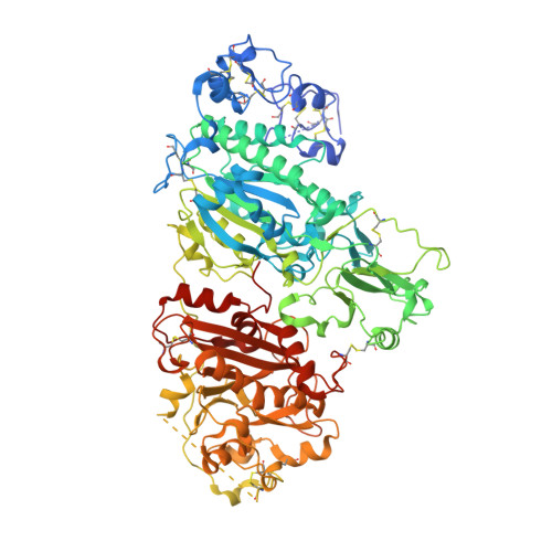
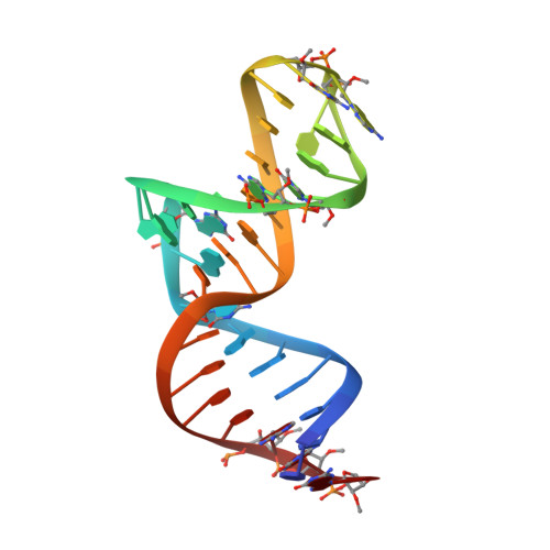
Structural basis for specific inhibition of Autotaxin by a DNA aptamer

Kato, K.Ikeda, H.Miyakawa, S.Futakawa, S.Nonaka, Y.Fujiwara, M.Okudaira, S.Kano, K.Aoki, J.Morita, J.Ishitani, R.Nishimasu, H.Nakamura, Y.Nureki, O.

(2016) Nat Struct Mol Biol 23: 395-401

  • DOI: https://doi.org/10.1038/nsmb.3200
  • Primary Citation Related Structures: 
    5HRT

  • PubMed Abstract: 

    ATX is a plasma lysophospholipase D that hydrolyzes lysophosphatidylcholine (LPC) and produces lysophosphatidic acid. To date, no ATX-inhibition-mediated treatment strategies for human diseases have been established. Here, we report anti-ATX DNA aptamers that inhibit ATX with high specificity and efficacy. We solved the crystal structure of ATX in complex with the anti-ATX aptamer RB011, at 2.0-Å resolution. RB011 binds in the vicinity of the active site through base-specific interactions, thus preventing the access of the choline moiety of LPC substrates. Using the structural information, we developed the modified anti-ATX DNA aptamer RB014, which exhibited in vivo efficacy in a bleomycin-induced pulmonary fibrosis mouse model. Our findings reveal the structural basis for the specific inhibition of ATX by the anti-ATX aptamer and highlight the therapeutic potential of anti-ATX aptamers for the treatment of human diseases, such as pulmonary fibrosis.


  • Organizational Affiliation
    • Department of Biological Sciences, Graduate School of Science, University of Tokyo, Tokyo, Japan.

Macromolecule Content 

  • Total Structure Weight: 109.7 kDa 
  • Atom Count: 7,677 
  • Modeled Residue Count: 820 
  • Deposited Residue Count: 865 
  • Unique protein chains: 1
  • Unique nucleic acid chains: 1

Macromolecules


Find similar proteins by:|  3D Structure
Entity ID: 1
MoleculeChains  Sequence LengthOrganismDetailsImage
Ectonucleotide pyrophosphatase/phosphodiesterase family member 2831Mus musculusMutation(s): 0 
Gene Names: Enpp2Npps2Pdnp2
EC: 3.1.4.39 (PDB Primary Data), 3.1.4.4 (UniProt)
UniProt
Find proteins for Q9R1E6 (Mus musculus)
Explore Q9R1E6 
Go to UniProtKB:  Q9R1E6
Entity Groups
Sequence Clusters30% Identity50% Identity70% Identity90% Identity95% Identity100% Identity
UniProt GroupQ9R1E6
Glycosylation
Glycosylation Sites: 2Go to GlyGen: Q9R1E6-1
Sequence Annotations
Expand
Reference Sequence
Find similar nucleic acids by:  (by identity cutoff) 
Entity ID: 2
MoleculeChains LengthOrganismImage
modified DNA (34-MER)34Homo sapiens
Sequence Annotations
Expand
Reference Sequence

Oligosaccharides

Help  
Entity ID: 3
MoleculeChains Length2D Diagram GlycosylationD Interactions
alpha-D-mannopyranose-(1-2)-alpha-D-mannopyranose-(1-3)-alpha-D-mannopyranose-(1-6)-[alpha-D-mannopyranose-(1-3)]beta-D-mannopyranose-(1-4)-2-acetamido-2-deoxy-beta-D-glucopyranose-(1-4)-2-acetamido-2-deoxy-beta-D-glucopyranose
C
7N-Glycosylation
Glycosylation Resources
GlyTouCan: G16404YH
GlyCosmos: G16404YH
GlyGen: G16404YH
Entity ID: 4
MoleculeChains Length2D Diagram GlycosylationD Interactions
alpha-D-mannopyranose-(1-3)-beta-D-mannopyranose-(1-4)-2-acetamido-2-deoxy-beta-D-glucopyranose-(1-4)-2-acetamido-2-deoxy-beta-D-glucopyranose
D
4N-Glycosylation
Glycosylation Resources
GlyTouCan: G81315DD
GlyCosmos: G81315DD
GlyGen: G81315DD

Small Molecules

Ligands 6 Unique
IDChains Name / Formula / InChI Key2D Diagram3D Interactions
GOL

Query on GOL


Download:Ideal Coordinates CCD File
K [auth A]
L [auth A]
M [auth A]
N [auth A]
O [auth A]
K [auth A],
L [auth A],
M [auth A],
N [auth A],
O [auth A],
P [auth A],
Q [auth A],
R [auth A],
S [auth A],
T [auth A]
GLYCEROL
C3 H8 O3
PEDCQBHIVMGVHV-UHFFFAOYSA-N
ZN

Query on ZN


Download:Ideal Coordinates CCD File
E [auth A],
F [auth A]
ZINC ION
Zn
PTFCDOFLOPIGGS-UHFFFAOYSA-N
CA

Query on CA


Download:Ideal Coordinates CCD File
G [auth A],
U [auth B]
CALCIUM ION
Ca
BHPQYMZQTOCNFJ-UHFFFAOYSA-N
K

Query on K


Download:Ideal Coordinates CCD File
J [auth A]POTASSIUM ION
K
NPYPAHLBTDXSSS-UHFFFAOYSA-N
CL

Query on CL


Download:Ideal Coordinates CCD File
I [auth A]CHLORIDE ION
Cl
VEXZGXHMUGYJMC-UHFFFAOYSA-M
NA

Query on NA


Download:Ideal Coordinates CCD File
H [auth A]SODIUM ION
Na
FKNQFGJONOIPTF-UHFFFAOYSA-N

Experimental Data & Validation

Experimental Data

  • Method: X-RAY DIFFRACTION
  • Resolution: 2.00 Å
  • R-Value Free:  0.202 (Depositor), 0.203 (DCC) 
  • R-Value Work:  0.164 (Depositor), 0.166 (DCC) 
  • R-Value Observed: 0.165 (Depositor) 
Space Group: C 2 2 21
Unit Cell:
Length ( Å )Angle ( ˚ )
a = 120.062α = 90
b = 208.757β = 90
c = 90.036γ = 90
Software Package:
Software NamePurpose
PHENIXrefinement
XDSdata processing
MOLREPphasing
Cootmodel building

Structure Validation

View Full Validation Report


Entry History 

Deposition Data

Revision History  (Full details and data files)

  • Version 1.0: 2016-04-06
    Type: Initial release
  • Version 1.1: 2016-05-25
    Changes: Database references
  • Version 1.2: 2020-02-19
    Changes: Data collection, Derived calculations
  • Version 2.0: 2020-07-29
    Type: Remediation
    Reason: Carbohydrate remediation
    Changes: Atomic model, Data collection, Derived calculations, Structure summary
  • Version 2.1: 2024-11-06
    Changes: Data collection, Database references, Derived calculations, Structure summary