A microbial sensor for organophosphate hydrolysis exploiting an engineered specificity switch in a transcription factor.
Jha, R.K., Kern, T.L., Kim, Y., Tesar, C., Jedrzejczak, R., Joachimiak, A., Strauss, C.E.(2016) Nucleic Acids Res 44: 8490-8500
- PubMed: 27536006 Search on PubMedSearch on PubMed Central
- DOI: https://doi.org/10.1093/nar/gkw687
- Primary Citation Related Structures:
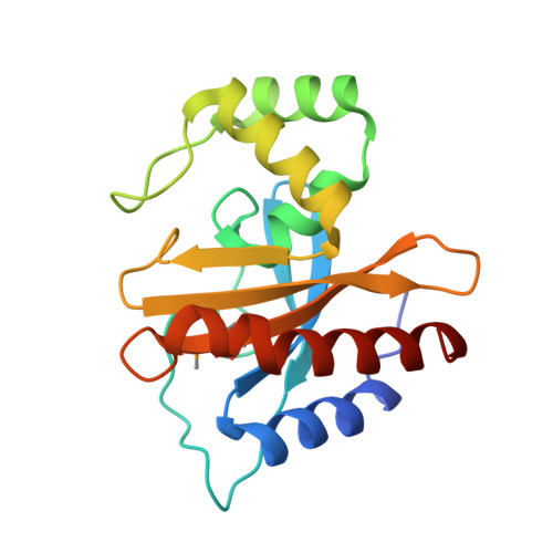
5HPF, 5HPI - PubMed Abstract:
A whole-cell biosensor utilizing a transcription factor (TF) is an effective tool for sensitive and selective detection of specialty chemicals or anthropogenic molecules, but requires access to an expanded repertoire of TFs. Using homology modeling and ligand docking for binding pocket identification, assisted by conservative mutations in the pocket, we engineered a novel specificity in an Acinetobacter TF, PobR, to 'sense' a chemical p-nitrophenol (pNP) and measured the response via a fluorescent protein reporter expressed from a PobR promoter. Out of 10(7) variants of PobR, four were active when dosed with pNP, with two mutants showing a specificity switch from the native effector 4-hydroxybenzoate (4HB). One of the mutants, pNPmut1 was then used to create a smart microbial cell responding to pNP production from hydrolysis of an insecticide, paraoxon, in a coupled assay involving phosphotriesterase (PTE) enzyme expressed from a separate promoter. We show the fluorescence of the cells correlated with the catalytic efficiency of the PTE variant expressed in each cell. High selectivity between similar molecules (4HB versus pNP), high sensitivity for pNP detection (∼2 μM) and agreement of apo- and holo-structures of PobR scaffold with predetermined computational models are other significant results presented in this work.
- Bioscience Division, PO Box 1663, Los Alamos National Laboratory, Los Alamos NM 87545, USA cems@lanl.gov.
Organizational Affiliation: