Structures of parasite calreticulins provide insights into their flexibility and dual carbohydrate/peptide-binding properties.
Moreau, C., Cioci, G., Iannello, M., Laffly, E., Chouquet, A., Ferreira, A., Thielens, N.M., Gaboriaud, C.(2016) IUCrJ 3: 408-419
- PubMed: 27840680 Search on PubMedSearch on PubMed Central
- DOI: https://doi.org/10.1107/S2052252516012847
- Primary Citation Related Structures:
5HCA, 5HCB, 5HCF, 5LK5 - PubMed Abstract:
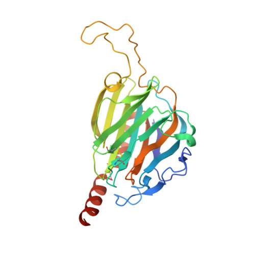
Calreticulin (CRT) is a multifaceted protein, initially discovered as an endoplasmic reticulum (ER) chaperone protein, that is essential in calcium metabolism. Various implications in cancer, early development and immunology have been discovered more recently for CRT, as well as its role as a dominant 'eat-me' prophagocytic signal. Intriguingly, cell-surface exposure/secretion of CRT is among the infective strategies used by parasites such as Trypanosoma cruzi , Entamoeba histolytica , Taenia solium , Leishmania donovani and Schistosoma mansoni . Because of the inherent flexibility of CRTs, their analysis by X-ray crystallography requires the design of recombinant constructs suitable for crystallization, and thus only the structures of two very similar mammalian CRT lectin domains are known. With the X-ray structures of two distant parasite CRTs, insights into species structural determinants that might be harnessed to fight against the parasites without affecting the functions of the host CRT are now provided. Moreover, although the hypothesis that CRT can exhibit both open and closed conformations has been proposed in relation to its chaperone function, only the open conformation has so far been observed in crystal structures. The first evidence is now provided of a complex conformational transition with the junction reoriented towards P-domain closure. SAXS experiments also provided additional information about the flexibility of T. cruzi CRT in solution, thus complementing crystallographic data on the open conformation. Finally, regarding the conserved lectin-domain structure and chaperone function, evidence is provided of its dual carbohydrate/protein specificity and a new scheme is proposed to interpret such unusual substrate-binding properties. These fascinating features are fully consistent with previous experimental observations, as discussed considering the broad spectrum of CRT sequence conservations and differences.
- Institut de Biologie Structurale (IBS), Université Grenoble Alpes, CEA, CNRS , 38044 Grenoble, France.
Organizational Affiliation: