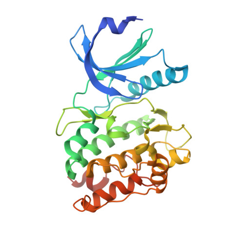
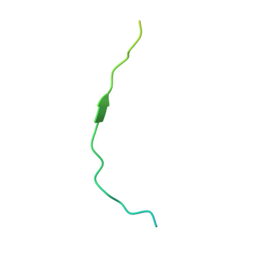
5H9B

Drosophila CaMKII-wt in complex with a fragment of the Eag potassium channel and Mg2+/AMPPN


Experimental Data Snapshot

  • Method: X-RAY DIFFRACTION
  • Resolution: 2.25 Å
  • R-Value Free: 0.222 
  • R-Value Work: 0.203 
  • R-Value Observed: 0.204 

wwPDB Validation   3D Report Full Report


Ligand Structure Quality Assessment 


This is version 1.1 of the entry. See complete history


Literature

The Interaction between the Drosophila EAG Potassium Channel and the Protein Kinase CaMKII Involves an Extensive Interface at the Active Site of the Kinase.

Castro-Rodrigues, A.F.Zhao, Y.Fonseca, F.Gabant, G.Cadene, M.Robertson, G.A.Morais-Cabral, J.H.

(2018) J Mol Biol 430: 5029-5049

  • DOI: https://doi.org/10.1016/j.jmb.2018.10.015
  • Primary Citation of Related Structures:  
    5FG8, 5H9B, 5HU3

  • PubMed Abstract: 

    The Drosophila EAG (dEAG) potassium channel is the founding member of the superfamily of KNCH channels, which are involved in cardiac repolarization, neuronal excitability and cellular proliferation. In flies, dEAG is involved in regulation of neuron firing and assembles with CaMKII to form a complex implicated in memory formation. We have characterized the interaction between the kinase domain of CaMKII and a 53-residue fragment of the dEAG channel that includes a canonical CaMKII recognition sequence. Crystal structures together with biochemical/biophysical analysis show a substrate-kinase complex with an unusually tight and extensive interface that appears to be strengthened by phosphorylation of the channel fragment. Electrophysiological recordings show that catalytically active CaMKII is required to observe active dEAG channels. A previously identified phosphorylation site in the recognition sequence is not the substrate for this crucial kinase activity, but rather contributes importantly to the tight interaction of the kinase with the channel. The available data suggest that the dEAG channel is a docking platform for the kinase and that phosphorylation of the channel's kinase recognition sequence modulates the strength of the interaction between the channel and the kinase.


  • Organizational Affiliation

    IBMC, Instituto de Biologia Molecular e Celular, Universidade do Porto, Porto, Portugal; i3S, Instituto de Investigação e Inovação em Saúde, Universidade do Porto, Porto, Portugal.


Macromolecules
Find similar proteins by:  (by identity cutoff)  |  3D Structure
Entity ID: 1
MoleculeChains Sequence LengthOrganismDetailsImage
Calcium/calmodulin-dependent protein kinase II, isoform C285Drosophila melanogasterMutation(s): 0 
Gene Names: CaMKIICaMKII-RACG18069Dmel_CG18069
EC: 2.7.11 (PDB Primary Data), 2.7.11.17 (PDB Primary Data)
UniProt
Find proteins for Q00168 (Drosophila melanogaster)
Explore Q00168 
Go to UniProtKB:  Q00168
Entity Groups  
Sequence Clusters30% Identity50% Identity70% Identity90% Identity95% Identity100% Identity
UniProt GroupQ00168
Sequence Annotations
Expand
  • Reference Sequence
Find similar proteins by:  (by identity cutoff)  |  3D Structure
Entity ID: 2
MoleculeChains Sequence LengthOrganismDetailsImage
Potassium voltage-gated channel protein eag54Drosophila melanogasterMutation(s): 0 
Gene Names: eagCG10952
UniProt
Find proteins for Q02280 (Drosophila melanogaster)
Explore Q02280 
Go to UniProtKB:  Q02280
Entity Groups  
Sequence Clusters30% Identity50% Identity70% Identity90% Identity95% Identity100% Identity
UniProt GroupQ02280
Sequence Annotations
Expand
  • Reference Sequence
Experimental Data & Validation

Experimental Data

  • Method: X-RAY DIFFRACTION
  • Resolution: 2.25 Å
  • R-Value Free: 0.222 
  • R-Value Work: 0.203 
  • R-Value Observed: 0.204 
  • Space Group: P 1 21 1
Unit Cell:
Length ( Å )Angle ( ˚ )
a = 35.823α = 90
b = 59.127β = 99.02
c = 70.339γ = 90
Software Package:
Software NamePurpose
SCALAdata scaling
PHENIXrefinement
PDB_EXTRACTdata extraction
XDSdata reduction
REFMACphasing

Structure Validation

View Full Validation Report


Ligand Structure Quality Assessment 


Entry History 

Deposition Data

Revision History  (Full details and data files)

  • Version 1.0: 2017-01-11
    Type: Initial release
  • Version 1.1: 2019-11-13
    Changes: Database references