5GXY

Crystal structure of endoglucanase CelQ from Clostridium thermocellum complexed with cellobiose and Tris


Experimental Data Snapshot

  • Method: X-RAY DIFFRACTION
  • Resolution: 1.70 Å
  • R-Value Free: 0.187 
  • R-Value Work: 0.160 
  • R-Value Observed: 0.161 

wwPDB Validation   3D Report Full Report


This is version 2.1 of the entry. See complete history


Literature

Crystal Structures of the C-Terminally Truncated Endoglucanase Cel9Q from Clostridium thermocellum Complexed with Cellodextrins and Tris.

Jeng, W.Y.Liu, C.I.Lu, T.J.Lin, H.J.Wang, N.C.Wang, A.H.

(2019) Chembiochem 20: 295-307

  • DOI: https://doi.org/10.1002/cbic.201800789
  • Primary Citation of Related Structures:  
    5GXX, 5GXY, 5GXZ, 5GY0, 5GY1

  • PubMed Abstract: 

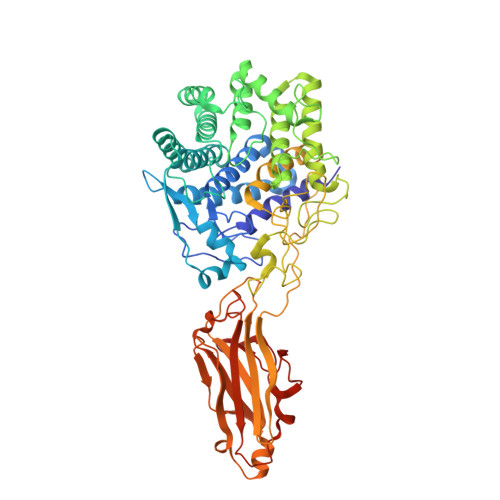
    Endoglucanase CtCel9Q is one of the enzyme components of the cellulosome, which is an active cellulase system in the thermophile Clostridium thermocellum. The precursor form of CtCel9Q comprises a signal peptide, a glycoside hydrolase family 9 catalytic domain, a type 3c carbohydrate-binding module (CBM), and a type I dockerin domain. Here, we report the crystal structures of C-terminally truncated CtCel9Q (CtCel9QΔc) complexed with Tris, Tris+cellobiose, cellobiose+cellotriose, cellotriose, and cellotetraose at resolutions of 1.50, 1.70, 2.05, 2.05 and 1.75 Å, respectively. CtCel9QΔc forms a V-shaped homodimer through residues Lys529-Glu542 on the type 3c CBM, which pairs two β-strands (β4 and β5 of the CBM). In addition, a disulfide bond was formed between the two Cys535 residues of the protein monomers in the asymmetric unit. The structures allow the identification of four minus (-) subsites and two plus (+) subsites; this is important for further understanding the structural basis of cellulose binding and hydrolysis. In the oligosaccharide-free and cellobiose-bound CtCel9QΔc structures, a Tris molecule was found to be bound to three catalytic residues of CtCel9Q and occupied subsite -1 of the CtCel9Q active-site cleft. Moreover, the enzyme activity assay in the presence of 100 mm Tris showed that the Tris almost completely suppressed CtCel9Q hydrolase activity.


  • Organizational Affiliation

    University Center for Bioscience and Biotechnology, National Cheng Kung University, 1 University Road, Tainan, 701, Taiwan.


Macromolecules
Find similar proteins by:  (by identity cutoff)  |  3D Structure
Entity ID: 1
MoleculeChains Sequence LengthOrganismDetailsImage
Glucanase
A, B
610Acetivibrio thermocellusMutation(s): 1 
Gene Names: celQ
EC: 3.2.1
UniProt
Find proteins for Q9AJF8 (Acetivibrio thermocellus)
Explore Q9AJF8 
Go to UniProtKB:  Q9AJF8
Entity Groups  
Sequence Clusters30% Identity50% Identity70% Identity90% Identity95% Identity100% Identity
UniProt GroupQ9AJF8
Sequence Annotations
Expand
  • Reference Sequence
Oligosaccharides

Help

Entity ID: 2
MoleculeChains Length2D Diagram Glycosylation3D Interactions
beta-D-glucopyranose-(1-4)-beta-D-glucopyranose
C, D
2N/A
Glycosylation Resources
GlyTouCan:  G84824ZO
GlyCosmos:  G84824ZO
Small Molecules
Ligands 5 Unique
IDChains Name / Formula / InChI Key2D Diagram3D Interactions
7PE
Query on 7PE

Download Ideal Coordinates CCD File 
Z [auth B]2-(2-(2-(2-(2-(2-ETHOXYETHOXY)ETHOXY)ETHOXY)ETHOXY)ETHOXY)ETHANOL
C14 H30 O7
UKXKPKBTMYNOFS-UHFFFAOYSA-N
TRS
Query on TRS

Download Ideal Coordinates CCD File 
O [auth A],
Y [auth B]
2-AMINO-2-HYDROXYMETHYL-PROPANE-1,3-DIOL
C4 H12 N O3
LENZDBCJOHFCAS-UHFFFAOYSA-O
BR
Query on BR

Download Ideal Coordinates CCD File 
L [auth A]
M [auth A]
N [auth A]
R [auth B]
W [auth B]
L [auth A],
M [auth A],
N [auth A],
R [auth B],
W [auth B],
X [auth B]
BROMIDE ION
Br
CPELXLSAUQHCOX-UHFFFAOYSA-M
CA
Query on CA

Download Ideal Coordinates CCD File 
E [auth A],
F [auth A],
P [auth B],
Q [auth B]
CALCIUM ION
Ca
BHPQYMZQTOCNFJ-UHFFFAOYSA-N
CL
Query on CL

Download Ideal Coordinates CCD File 
G [auth A]
H [auth A]
I [auth A]
J [auth A]
K [auth A]
G [auth A],
H [auth A],
I [auth A],
J [auth A],
K [auth A],
S [auth B],
T [auth B],
U [auth B],
V [auth B]
CHLORIDE ION
Cl
VEXZGXHMUGYJMC-UHFFFAOYSA-M
Biologically Interesting Molecules (External Reference) 1 Unique
Experimental Data & Validation

Experimental Data

  • Method: X-RAY DIFFRACTION
  • Resolution: 1.70 Å
  • R-Value Free: 0.187 
  • R-Value Work: 0.160 
  • R-Value Observed: 0.161 
  • Space Group: P 21 21 21
Unit Cell:
Length ( Å )Angle ( ˚ )
a = 108.216α = 90
b = 108.822β = 90
c = 139.816γ = 90
Software Package:
Software NamePurpose
REFMACrefinement
HKL-2000data reduction
HKL-2000data scaling
PHASERphasing

Structure Validation

View Full Validation Report


Entry History & Funding Information

Deposition Data


Funding OrganizationLocationGrant Number
Ministry of Science and Technology of TaiwanTaiwanNSC 102-2311-B-006-006-MY3
Ministry of Science and Technology of TaiwanTaiwanNSC 99-3113-B-001-001
Ministry of Science and Technology of TaiwanTaiwanMOST105-0210-01-12-01

Revision History  (Full details and data files)

  • Version 1.0: 2017-09-27
    Type: Initial release
  • Version 1.1: 2019-04-10
    Changes: Data collection, Database references
  • Version 2.0: 2020-07-29
    Type: Remediation
    Reason: Carbohydrate remediation
    Changes: Advisory, Atomic model, Data collection, Derived calculations, Structure summary
  • Version 2.1: 2023-11-08
    Changes: Data collection, Database references, Derived calculations, Refinement description, Structure summary