5GRF

Crystal structure of the alpha gamma mutant (gamma-K151A) of human IDH3 in complex with Mg(2+), citrate and ADP

  • Classification: OXIDOREDUCTASE
  • Organism(s): Homo sapiens
  • Expression System: Escherichia coli BL21(DE3)
  • Mutation(s): Yes 

  • Deposited: 2016-08-11 Released: 2017-02-15 
  • Deposition Author(s): Ma, T., Ding, J.
  • Funding Organization(s): the National Natural Science Foundation of China, the Ministry of Science and Technology of China, the Chinese Academy of Sciences

Experimental Data Snapshot

  • Method: X-RAY DIFFRACTION
  • Resolution: 2.50 Å
  • R-Value Free: 0.247 
  • R-Value Work: 0.207 
  • R-Value Observed: 0.209 

wwPDB Validation   3D Report Full Report


Ligand Structure Quality Assessment 


This is version 1.2 of the entry. See complete history


Literature

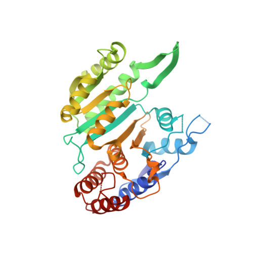
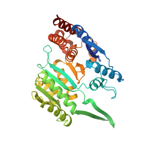
Molecular mechanism of the allosteric regulation of the alpha gamma heterodimer of human NAD-dependent isocitrate dehydrogenase.

Ma, T.Peng, Y.Huang, W.Ding, J.

(2017) Sci Rep 7: 40921-40921

  • DOI: https://doi.org/10.1038/srep40921
  • Primary Citation of Related Structures:  
    5GRE, 5GRF, 5GRH, 5GRI, 5GRL

  • PubMed Abstract: 

    Human NAD-dependent isocitrate dehydrogenase catalyzes the decarboxylation of isocitrate (ICT) into α-ketoglutarate in the Krebs cycle. It exists as the α 2 βγ heterotetramer composed of the αβ and αγ heterodimers. Previously, we have demonstrated biochemically that the α 2 βγ heterotetramer and αγ heterodimer can be allosterically activated by citrate (CIT) and ADP. In this work, we report the crystal structures of the αγ heterodimer with the γ subunit bound without or with different activators. Structural analyses show that CIT, ADP and Mg 2+ bind adjacent to each other at the allosteric site. The CIT binding induces conformational changes at the allosteric site, which are transmitted to the active site through the heterodimer interface, leading to stabilization of the ICT binding at the active site and thus activation of the enzyme. The ADP binding induces no further conformational changes but enhances the CIT binding through Mg 2+ -mediated interactions, yielding a synergistic activation effect. ICT can also bind to the CIT-binding subsite, which induces similar conformational changes but exhibits a weaker activation effect. The functional roles of the key residues are verified by mutagenesis, kinetic and structural studies. Our structural and functional data together reveal the molecular mechanism of the allosteric regulation of the αγ heterodimer.


  • Organizational Affiliation

    National Center for Protein Science Shanghai, State Key Laboratory of Molecular Biology, Center for Excellence in Molecular Cell Science, Institute of Biochemistry and Cell Biology, Shanghai Institutes for Biological Sciences, Chinese Academy of Sciences, 320 Yueyang Road, Shanghai 200031, China.


Macromolecules
Find similar proteins by:  (by identity cutoff)  |  3D Structure
Entity ID: 1
MoleculeChains Sequence LengthOrganismDetailsImage
Isocitrate dehydrogenase [NAD] subunit alpha, mitochondrial339Homo sapiensMutation(s): 0 
Gene Names: IDH3A
EC: 1.1.1.41
UniProt & NIH Common Fund Data Resources
Find proteins for P50213 (Homo sapiens)
Explore P50213 
Go to UniProtKB:  P50213
PHAROS:  P50213
GTEx:  ENSG00000166411 
Entity Groups  
Sequence Clusters30% Identity50% Identity70% Identity90% Identity95% Identity100% Identity
UniProt GroupP50213
Sequence Annotations
Expand
  • Reference Sequence
Find similar proteins by:  (by identity cutoff)  |  3D Structure
Entity ID: 2
MoleculeChains Sequence LengthOrganismDetailsImage
Isocitrate dehydrogenase [NAD] subunit gamma, mitochondrial354Homo sapiensMutation(s): 1 
Gene Names: IDH3G
EC: 1.1.1.41
UniProt & NIH Common Fund Data Resources
Find proteins for P51553 (Homo sapiens)
Explore P51553 
Go to UniProtKB:  P51553
PHAROS:  P51553
GTEx:  ENSG00000067829 
Entity Groups  
Sequence Clusters30% Identity50% Identity70% Identity90% Identity95% Identity100% Identity
UniProt GroupP51553
Sequence Annotations
Expand
  • Reference Sequence
Experimental Data & Validation

Experimental Data

  • Method: X-RAY DIFFRACTION
  • Resolution: 2.50 Å
  • R-Value Free: 0.247 
  • R-Value Work: 0.207 
  • R-Value Observed: 0.209 
  • Space Group: P 31 2 1
Unit Cell:
Length ( Å )Angle ( ˚ )
a = 114.336α = 90
b = 114.336β = 90
c = 143.901γ = 120
Software Package:
Software NamePurpose
REFMACrefinement
PDB_EXTRACTdata extraction
PHASERphasing

Structure Validation

View Full Validation Report


Ligand Structure Quality Assessment 


Entry History & Funding Information

Deposition Data


Funding OrganizationLocationGrant Number
the National Natural Science Foundation of ChinaChina31170690
the Ministry of Science and Technology of ChinaChina2011CB911102
the Chinese Academy of SciencesChinaXDB08010302

Revision History  (Full details and data files)

  • Version 1.0: 2017-02-15
    Type: Initial release
  • Version 1.1: 2017-10-04
    Changes: Data collection
  • Version 1.2: 2023-11-08
    Changes: Data collection, Database references, Refinement description