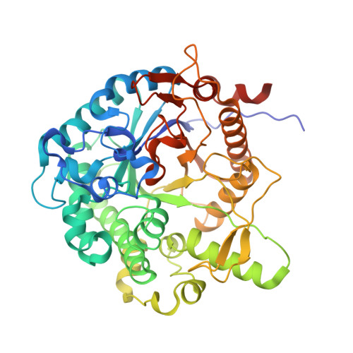
Structures of a glucose-tolerant beta-glucosidase provide insights into its mechanism.
Pang, P., Cao, L.C., Liu, Y.H., Xie, W., Wang, Z.(2017) J Struct Biol 198: 154-162
- PubMed: 28189793 Search on PubMed
- DOI: https://doi.org/10.1016/j.jsb.2017.02.001
- Primary Citation Related Structures:
5GNX, 5GNY, 5GNZ - PubMed Abstract:
Cellulose can be converted to ethanol via the fermentation of glucose, which is considered as a promising green alternative for transportation fuels. The conversion of cellulose to glucose needs three enzymes, in which β-glucosidase (BGL) plays an essential role. However, BGL is inhibited by its own product glucose, greatly limiting its applications in industry. We previously obtained a novel BGL named Bgl6 with a high glucose tolerance. Further engineering through random mutagenesis produced a triple mutant M3 with improved thermostability. This enzyme shows promising properties for wide applications but the structural basis of the unusual properties of Bgl6 is not clear. In this study, we determined the crystal structures of Bgl6 and variants at high resolution, which provide insights into its glucose-tolerant mechanism and thermostability. Particularly, Bgl6 forms an extra channel that could be used as a secondary binding site for glucose, which may contribute to glucose tolerance. Additionally, the triple mutations could strengthen the hydrophobic interactions within the enzyme and may be responsible for the enhanced thermostability exhibited by M3, which was further confirmed by dynamic light scattering data. Lastly, structural comparison to other orthologs allows us to formulate new strategies on how to improve the catalytic efficiency of Bgl6.
- School of Pharmaceutical Sciences, The Sun Yat-Sen University, 132 E. Circle Rd. University City, Guangzhou, Guangdong 510006, People's Republic of China; Center for Cellular & Structural Biology, The Sun Yat-Sen University, 132 E. Circle Rd., University City, Guangzhou, Guangdong 510006, People's Republic of China.
Organizational Affiliation: