Structure and Polymannuronate Specificity of a Eukaryotic Member of Polysaccharide Lyase Family 14.
Qin, H.M., Miyakawa, T., Inoue, A., Nishiyama, R., Nakamura, A., Asano, A., Sawano, Y., Ojima, T., Tanokura, M.(2017) J Biol Chem 292: 2182-2190
- PubMed: 28011642 Search on PubMedSearch on PubMed Central
- DOI: https://doi.org/10.1074/jbc.M116.749929
- Primary Citation Related Structures:
5GMT - PubMed Abstract:
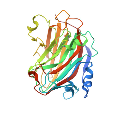
Alginate is an abundant algal polysaccharide, composed of β-d-mannuronate and its C5 epimer α-l-guluronate, that is a useful biomaterial in cell biology and tissue engineering, with applications in cancer and aging research. The alginate lyase (EC 4.2.2.3) from Aplysia kurodai , AkAly30, is a eukaryotic member of the polysaccharide lyase 14 (PL-14) family and degrades alginate by cleaving the glycosidic bond through a β-elimination reaction. Here, we present the structural basis for the substrate specificity, with a preference for polymannuronate, of AkAly30. The crystal structure of AkAly30 at a 1.77 Å resolution and the putative substrate-binding model show that the enzyme adopts a β-jelly roll fold at the core of the structure and that Lys-99, Tyr-140, and Tyr-142 form catalytic residues in the active site. Their arrangements allow the carboxyl group of mannuronate residues at subsite +1 to form ionic bonds with Lys-99. The coupled tyrosine forms a hydrogen bond network with the glycosidic bond, and the hydroxy group of Tyr-140 is located near the C5 atom of the mannuronate residue. These interactions could promote the β-elimination of the mannuronate residue at subsite +1. More interestingly, Gly-118 and the disulfide bond formed by Cys-115 and Cys-124 control the conformation of an active-site loop, which makes the space suitable for substrate entry into subsite -1. The cleavage efficiency of AkAly30 is enhanced relative to that of mutants lacking either Gly-118 or the Cys-115-Cys-124 disulfide bond. The putative binding model and mutagenesis studies provide a novel substrate recognition mode explaining the polymannuronate specificity of PL-14 alginate lyases.
- From the Department of Applied Biological Chemistry, Graduate School of Agricultural and Life Sciences, University of Tokyo, 1-1-1 Yayoi, Bunkyo-ku, Tokyo 113-8657, Japan.
Organizational Affiliation: