Heterogeneity in the Histidine-Brace Copper Coordination Sphere in Aa10 Lytic Polysaccharide Monooxygenases.
Chaplin, A.K., Wilson, M.T., Hough, M.A., Svistunenko, D.A., Hemsworth, G.R., Walton, P.H., Vijgenboom, E., Worrall, J.A.R.(2016) J Biological Chem 291: 12838
- PubMed: 27129229 Search on PubMedSearch on PubMed Central
- DOI: https://doi.org/10.1074/jbc.M116.722447
- Primary Citation Related Structures:
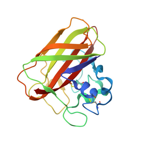
5FTZ - PubMed Abstract:
Copper-dependent lytic polysaccharide monooxygenases (LPMOs) are enzymes that oxidatively deconstruct polysaccharides. The active site copper in LPMOs is coordinated by a histidine-brace. This utilizes the amino group and side chain of the N-terminal His residue with the side chain of a second His residue to create a T-shaped arrangement of nitrogen ligands. We report a structural, kinetic, and thermodynamic appraisal of copper binding to the histidine-brace in an auxiliary activity family 10 (AA10) LPMO from Streptomyces lividans (SliLPMO10E). Unexpectedly, we discovered the existence of two apo-SliLPMO10E species in solution that can each bind copper at a single site with distinct kinetic and thermodynamic (exothermic and endothermic) properties. The experimental EPR spectrum of copper-bound SliLPMO10E requires the simulation of two different line shapes, implying two different copper-bound species, indicative of three and two nitrogen ligands coordinating the copper. Amino group coordination was probed through the creation of an N-terminal extension variant (SliLPMO10E-Ext). The kinetics and thermodynamics of copper binding to SliLPMO10E-Ext are in accord with copper binding to one of the apo-forms in the wild-type protein, suggesting that amino group coordination is absent in the two-nitrogen coordinate form of SliLPMO10E. Copper binding to SliLPMO10B was also investigated, and again it revealed the presence of two apo-forms with kinetics and stoichiometry of copper binding identical to that of SliLPMO10E. Our findings highlight that heterogeneity exists in the active site copper coordination sphere of LPMOs that may have implications for the mechanism of loading copper in the cell.
- From the School of Biological Sciences, University of Essex, Wivenhoe Park, Colchester CO4 3SQ, United Kingdom.
Organizational Affiliation: