5FOE

Crystal structure of the C. elegans Protein O-fucosyltransferase 2 (CePOFUT2) double mutant (R298K-R299K) in complex with GDP and the human TSR1 from thrombospondin 1


Experimental Data Snapshot

  • Method: X-RAY DIFFRACTION
  • Resolution: 1.98 Å
  • R-Value Free: 0.246 
  • R-Value Work: 0.200 
  • R-Value Observed: 0.202 

wwPDB Validation   3D Report Full Report


Ligand Structure Quality Assessment 


This is version 1.6 of the entry. See complete history


Literature

A Proactive Role of Water Molecules in Acceptor Recognition of Thrombospondin Type 1 Repeats by Protein-O-Fucosyltransferase 2

Valero-Gonzalez, J.Leonhard-Melief, C.Lira-Navarrete, E.Jimenez-Oses, G.Hernandez-Ruiz, C.Pallares, M.C.Yruela, I.Vasudevan, D.Lostao, A.Corzana, F.Takeuchi, H.Haltiwanger, R.S.Hurtado-Guerrero, R.

(2016) Nat Chem Biol 12: 240

  • DOI: https://doi.org/10.1038/nchembio.2019
  • Primary Citation of Related Structures:  
    5FOE

  • PubMed Abstract: 

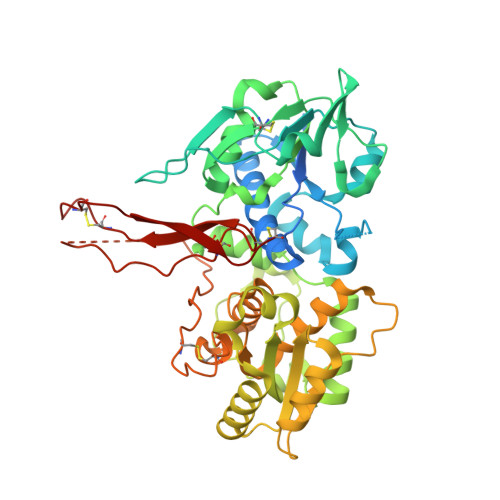
    Protein O-fucosyltransferase 2 (POFUT2) is an essential enzyme that fucosylates serine and threonine residues of folded thrombospondin type 1 repeats (TSRs). To date, the mechanism by which this enzyme recognizes very dissimilar TSRs has been unclear. By engineering a fusion protein, we report the crystal structure of Caenorhabditis elegans POFUT2 (CePOFUT2) in complex with GDP and human TSR1 that suggests an inverting mechanism for fucose transfer assisted by a catalytic base and shows that nearly half of the TSR1 is embraced by CePOFUT2. A small number of direct interactions and a large network of water molecules maintain the complex. Site-directed mutagenesis demonstrates that POFUT2 fucosylates threonine preferentially over serine and relies on folded TSRs containing the minimal consensus sequence C-X-X-S/T-C. Crystallographic and mutagenesis data, together with atomic-level simulations, uncover a binding mechanism by which POFUT2 promiscuously recognizes the structural fingerprint of poorly homologous TSRs through a dynamic network of water-mediated interactions.


  • Organizational Affiliation

    Institute for Biocomputation and Physics of Complex Systems (BIFI), University of Zaragoza, BIFI-IQFR (CSIC) Joint Unit, Mariano Esquillor s/n, Campus Rio Ebro, Zaragoza, Spain.


Macromolecules
Find similar proteins by:  (by identity cutoff)  |  3D Structure
Entity ID: 1
MoleculeChains Sequence LengthOrganismDetailsImage
GDP-fucose protein O-fucosyltransferase 2,Thrombospondin-1
A, B
500Caenorhabditis elegansHomo sapiens
This entity is chimeric
Mutation(s): 2 
Gene Names: pad-2K10G9.3THBS1TSPTSP1
EC: 2.4.1.221
UniProt & NIH Common Fund Data Resources
Find proteins for Q8WR51 (Caenorhabditis elegans)
Explore Q8WR51 
Go to UniProtKB:  Q8WR51
Find proteins for P07996 (Homo sapiens)
Explore P07996 
Go to UniProtKB:  P07996
PHAROS:  P07996
GTEx:  ENSG00000137801 
Entity Groups  
Sequence Clusters30% Identity50% Identity70% Identity90% Identity95% Identity100% Identity
UniProt GroupsP07996Q8WR51
Sequence Annotations
Expand
  • Reference Sequence
Small Molecules
Ligands 3 Unique
IDChains Name / Formula / InChI Key2D Diagram3D Interactions
GDP
Query on GDP

Download Ideal Coordinates CCD File 
D [auth A],
J [auth B]
GUANOSINE-5'-DIPHOSPHATE
C10 H15 N5 O11 P2
QGWNDRXFNXRZMB-UUOKFMHZSA-N
NAG
Query on NAG

Download Ideal Coordinates CCD File 
C [auth A],
K [auth B]
2-acetamido-2-deoxy-beta-D-glucopyranose
C8 H15 N O6
OVRNDRQMDRJTHS-FMDGEEDCSA-N
EDO
Query on EDO

Download Ideal Coordinates CCD File 
E [auth A]
F [auth A]
G [auth A]
H [auth A]
I [auth A]
E [auth A],
F [auth A],
G [auth A],
H [auth A],
I [auth A],
L [auth B],
M [auth B],
N [auth B],
O [auth B],
P [auth B]
1,2-ETHANEDIOL
C2 H6 O2
LYCAIKOWRPUZTN-UHFFFAOYSA-N
Experimental Data & Validation

Experimental Data

  • Method: X-RAY DIFFRACTION
  • Resolution: 1.98 Å
  • R-Value Free: 0.246 
  • R-Value Work: 0.200 
  • R-Value Observed: 0.202 
  • Space Group: P 1 21 1
Unit Cell:
Length ( Å )Angle ( ˚ )
a = 88.026α = 90
b = 67.447β = 116.07
c = 90.411γ = 90
Software Package:
Software NamePurpose
REFMACrefinement
XDSdata reduction
SCALEPACKdata scaling
PHASERphasing

Structure Validation

View Full Validation Report


Ligand Structure Quality Assessment 


Entry History 

Revision History  (Full details and data files)

  • Version 1.0: 2016-01-27
    Type: Initial release
  • Version 1.1: 2016-02-10
    Changes: Database references
  • Version 1.2: 2016-03-02
    Changes: Database references
  • Version 1.3: 2016-03-30
    Changes: Database references
  • Version 1.4: 2017-03-15
    Changes: Source and taxonomy
  • Version 1.5: 2020-07-29
    Type: Remediation
    Reason: Carbohydrate remediation
    Changes: Data collection, Derived calculations, Other, Structure summary
  • Version 1.6: 2024-01-10
    Changes: Data collection, Database references, Refinement description, Structure summary