5FMW | pdb_00005fmw

The poly-C9 component of the Complement Membrane Attack Complex


Experimental Data Snapshot

  • Method: ELECTRON MICROSCOPY
  • Resolution: 6.70 Å
  • Aggregation State: PARTICLE 
  • Reconstruction Method: SINGLE PARTICLE 

wwPDB Validation 3D Report Full Report

Validation slider image for 5FMW

This is version 1.2 of the entry. See complete history

Literature

Structure of the Poly-C9 Component of the Complement Membrane Attack Complex

Dudkina, N.V.Spicer, B.A.Reboul, C.F.Conroy, P.J.Lukoyanova, N.Elmlund, H.Law, R.H.P.Ekkel, S.M.Kondos, S.C.Goode, R.J.A.Ramm, G.Whisstock, J.C.Saibil, H.R.Dunstone, M.A.

(2016) Nat Commun 7: 10588

  • DOI: https://doi.org/10.1038/ncomms10588
  • Primary Citation Related Structures: 
    5FMW

  • PubMed Abstract: 

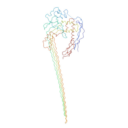
    The membrane attack complex (MAC)/perforin-like protein complement component 9 (C9) is the major component of the MAC, a multi-protein complex that forms pores in the membrane of target pathogens. In contrast to homologous proteins such as perforin and the cholesterol-dependent cytolysins (CDCs), all of which require the membrane for oligomerisation, C9 assembles directly onto the nascent MAC from solution. However, the molecular mechanism of MAC assembly remains to be understood. Here we present the 8 Å cryo-EM structure of a soluble form of the poly-C9 component of the MAC. These data reveal a 22-fold symmetrical arrangement of C9 molecules that yield an 88-strand pore-forming β-barrel. The N-terminal thrombospondin-1 (TSP1) domain forms an unexpectedly extensive part of the oligomerisation interface, thus likely facilitating solution-based assembly. These TSP1 interactions may also explain how additional C9 subunits can be recruited to the growing MAC subsequent to membrane insertion.


  • Organizational Affiliation
    • Department of Crystallography, Institute of Structural and Molecular Biology, Birkbeck College, London WC1E 7HX, UK.

Macromolecule Content 

  • Total Structure Weight: 1,266.82 kDa 
  • Atom Count: 10,582 
  • Modeled Residue Count: 10,582 
  • Deposited Residue Count: 11,132 
  • Unique protein chains: 1

Macromolecules

Find similar proteins by:|  3D Structure
Entity ID: 1
MoleculeChains  Sequence LengthOrganismDetailsImage
POLYC9
A, B, C, D, E
A, B, C, D, E, F, G, H, I, J, K, L, M, N, O, P, Q, R, S, T, U, V
506Homo sapiensMutation(s): 0 
Membrane Entity: Yes 
UniProt & NIH Common Fund Data Resources
Find proteins for P02748 (Homo sapiens)
Explore P02748 
Go to UniProtKB:  P02748
PHAROS:  P02748
GTEx:  ENSG00000113600 
Entity Groups
Sequence Clusters30% Identity50% Identity70% Identity90% Identity95% Identity100% Identity
UniProt GroupP02748
Sequence Annotations
Expand
Reference Sequence

Experimental Data & Validation

Experimental Data

  • Method: ELECTRON MICROSCOPY
  • Resolution: 6.70 Å
  • Aggregation State: PARTICLE 
  • Reconstruction Method: SINGLE PARTICLE 
EM Software:
TaskSoftware PackageVersion
RECONSTRUCTIONIMAGIC
RECONSTRUCTIONRELION

Structure Validation

View Full Validation Report


Entry History 

Revision History  (Full details and data files)

  • Version 1.0: 2016-02-17
    Type: Initial release
  • Version 1.1: 2017-08-02
    Changes: Data collection
  • Version 1.2: 2024-05-08
    Changes: Data collection, Database references