5FKK

TetR(D) N82A mutant in complex with anhydrotetracycline and magnesium


Experimental Data Snapshot

  • Method: X-RAY DIFFRACTION
  • Resolution: 1.75 Å
  • R-Value Free: 0.254 
  • R-Value Work: 0.207 
  • R-Value Observed: 0.209 

wwPDB Validation   3D Report Full Report


Ligand Structure Quality Assessment 


This is version 1.5 of the entry. See complete history


Literature

Modular Organisation of Inducer Recognition and Allostery in the Tetracycline Repressor

Werten, S.Schneider, J.Palm, G.J.Hinrichs, W.

(2016) FEBS J 283: 2102

  • DOI: https://doi.org/10.1111/febs.13723
  • Primary Citation of Related Structures:  
    5FKK, 5FKL, 5FKM, 5FKN, 5FKO

  • PubMed Abstract: 

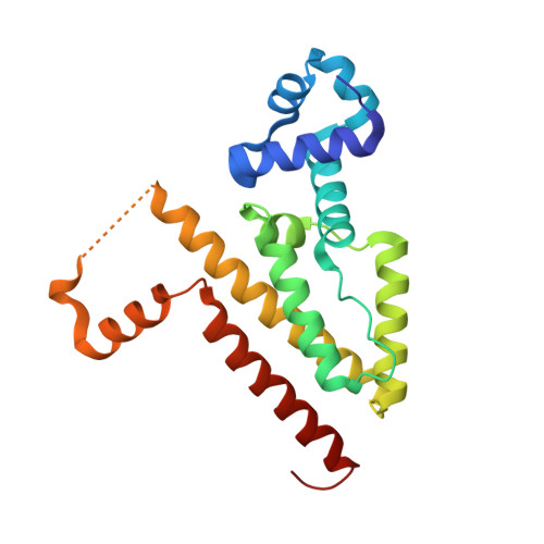
    Induction of the tetracycline repressor (TetR) results from antibiotic-dependent changes in the relative positioning of the DNA-binding domains within the promoter-associated repressor dimer, but the key determinants of this allosteric effect remain poorly characterised. Intriguingly, previous mutational analyses of the tetracycline-interacting site revealed a lack of correlation between residual affinity and induction propensity, suggesting that some of the residues in contact with the antibiotic primarily act in ligand recognition and retention, whereas others are required to transmit the allosteric signal. Here, we provide a structural basis for these observations via crystallographic analysis of the point mutants N82A, H100A, T103A and E147A in complex with the inducer 5a,6-anhydrotetracycline. In conjunction with the available functional data, the four structures demonstrate that a trigger-like movement of the region between helices α6 and α7 towards and into the binding site plays a decisive role in the intramolecular communication process. In sharp contrast, residues lining the binding cavity proper have little or no influence on the allosteric mechanism as such. This nearly complete physical separation of ligand recognition and allostery will have allowed diverging TetR-like repressors to bind novel effectors while the existing induction mechanism remained intact. Consequently, the modularity described here may have been a key factor in the evolutionary success of the widespread and highly diversified repressor class.


  • Organizational Affiliation

    Department of Molecular Structural Biology, Institute for Biochemistry, University of Greifswald, Germany.


Macromolecules
Find similar proteins by:  (by identity cutoff)  |  3D Structure
Entity ID: 1
MoleculeChains Sequence LengthOrganismDetailsImage
TETRACYCLINE REPRESSOR, CLASS D
A, B
207Escherichia coliMutation(s): 1 
UniProt
Find proteins for P0ACT4 (Escherichia coli)
Explore P0ACT4 
Go to UniProtKB:  P0ACT4
Entity Groups  
Sequence Clusters30% Identity50% Identity70% Identity90% Identity95% Identity100% Identity
UniProt GroupP0ACT4
Sequence Annotations
Expand
  • Reference Sequence
Small Molecules
Ligands 6 Unique
IDChains Name / Formula / InChI Key2D Diagram3D Interactions
TDC
Query on TDC

Download Ideal Coordinates CCD File 
I [auth B]5A,6-ANHYDROTETRACYCLINE
C22 H22 N2 O7
CXCVEERYMJZMMM-DOCRCCHOSA-N
PG4
Query on PG4

Download Ideal Coordinates CCD File 
C [auth A]TETRAETHYLENE GLYCOL
C8 H18 O5
UWHCKJMYHZGTIT-UHFFFAOYSA-N
PGE
Query on PGE

Download Ideal Coordinates CCD File 
P [auth B]TRIETHYLENE GLYCOL
C6 H14 O4
ZIBGPFATKBEMQZ-UHFFFAOYSA-N
SO4
Query on SO4

Download Ideal Coordinates CCD File 
D [auth A]SULFATE ION
O4 S
QAOWNCQODCNURD-UHFFFAOYSA-L
CL
Query on CL

Download Ideal Coordinates CCD File 
E [auth A]
F [auth A]
G [auth A]
H [auth A]
K [auth B]
E [auth A],
F [auth A],
G [auth A],
H [auth A],
K [auth B],
L [auth B],
M [auth B],
N [auth B],
O [auth B]
CHLORIDE ION
Cl
VEXZGXHMUGYJMC-UHFFFAOYSA-M
MG
Query on MG

Download Ideal Coordinates CCD File 
J [auth B]MAGNESIUM ION
Mg
JLVVSXFLKOJNIY-UHFFFAOYSA-N
Experimental Data & Validation

Experimental Data

  • Method: X-RAY DIFFRACTION
  • Resolution: 1.75 Å
  • R-Value Free: 0.254 
  • R-Value Work: 0.207 
  • R-Value Observed: 0.209 
  • Space Group: P 43 21 2
Unit Cell:
Length ( Å )Angle ( ˚ )
a = 68.231α = 90
b = 68.231β = 90
c = 179.247γ = 90
Software Package:
Software NamePurpose
REFMACrefinement
HKL-2000data reduction
HKL-2000data scaling
PHASERphasing

Structure Validation

View Full Validation Report


Ligand Structure Quality Assessment 


Entry History 

Deposition Data

Revision History  (Full details and data files)

  • Version 1.0: 2016-04-06
    Type: Initial release
  • Version 1.1: 2016-04-13
    Changes: Database references
  • Version 1.2: 2016-06-15
    Changes: Database references
  • Version 1.3: 2019-05-08
    Changes: Data collection, Experimental preparation, Other
  • Version 1.4: 2019-07-17
    Changes: Data collection
  • Version 1.5: 2024-01-10
    Changes: Data collection, Database references, Derived calculations, Other, Refinement description