Efficient switching of mCherry fluorescence using chemical caging.
Cloin, B.M.C., De Zitter, E., Salas, D., Gielen, V., Folkers, G.E., Mikhaylova, M., Bergeler, M., Krajnik, B., Harvey, J., Hoogenraad, C.C., Van Meervelt, L., Dedecker, P., Kapitein, L.C.(2017) Proc Natl Acad Sci U S A 114: 7013-7018
- PubMed: 28630286 Search on PubMedSearch on PubMed Central
- DOI: https://doi.org/10.1073/pnas.1617280114
- Primary Citation Related Structures:
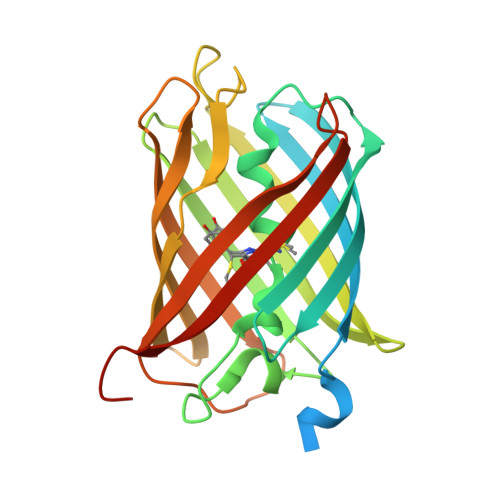
5FHV - PubMed Abstract:
Fluorophores with dynamic or controllable fluorescence emission have become essential tools for advanced imaging, such as superresolution imaging. These applications have driven the continuing development of photoactivatable or photoconvertible labels, including genetically encoded fluorescent proteins. These new probes work well but require the introduction of new labels that may interfere with the proper functioning of existing constructs and therefore require extensive functional characterization. In this work we show that the widely used red fluorescent protein mCherry can be brought to a purely chemically induced blue-fluorescent state by incubation with β-mercaptoethanol (βME). The molecules can be recovered to the red fluorescent state by washing out the βME or through irradiation with violet light, with up to 80% total recovery. We show that this can be used to perform single-molecule localization microscopy (SMLM) on cells expressing mCherry, which renders this approach applicable to a very wide range of existing constructs. We performed a detailed investigation of the mechanism underlying these dynamics, using X-ray crystallography, NMR spectroscopy, and ab initio quantum-mechanical calculations. We find that the βME-induced fluorescence quenching of mCherry occurs both via the direct addition of βME to the chromophore and through βME-mediated reduction of the chromophore. These results not only offer a strategy to expand SMLM imaging to a broad range of available biological models, but also present unique insights into the chemistry and functioning of a highly important class of fluorophores.
- Cell Biology, Department of Biology, Faculty of Science, Utrecht University, 3584 CH Utrecht, The Netherlands.
Organizational Affiliation: