Binding of Gd(3+) to the neuronal signalling protein calexcitin identifies an exchangeable Ca(2+)-binding site.
Chataigner, L., Guo, J., Erskine, P.T., Coker, A.R., Wood, S.P., Gombos, Z., Cooper, J.B.(2016) Acta Crystallogr F Struct Biol Commun 72: 276-281
- PubMed: 27050260 Search on PubMedSearch on PubMed Central
- DOI: https://doi.org/10.1107/S2053230X16003526
- Primary Citation Related Structures:
5F6T - PubMed Abstract:
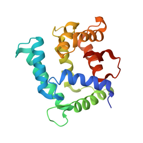
Calexcitin was first identified in the marine snail Hermissenda crassicornis as a neuronal-specific protein that becomes upregulated and phosphorylated in associative learning. Calexcitin possesses four EF-hand motifs, but only the first three (EF-1 to EF-3) are involved in binding metal ions. Past work has indicated that under physiological conditions EF-1 and EF-2 bind Mg(2+) and Ca(2+), while EF-3 is likely to bind only Ca(2+). The fourth EF-hand is nonfunctional owing to a lack of key metal-binding residues. The aim of this study was to use a crystallographic approach to determine which of the three metal-binding sites of calexcitin is most readily replaced by exogenous metal ions, potentially shedding light on which of the EF-hands play a `sensory' role in neuronal calcium signalling. By co-crystallizing recombinant calexcitin with equimolar Gd(3+) in the presence of trace Ca(2+), EF-1 was shown to become fully occupied by Gd(3+) ions, while the other two sites remain fully occupied by Ca(2+). The structure of the Gd(3+)-calexcitin complex has been refined to an R factor of 21.5% and an Rfree of 30.4% at 2.2 Å resolution. These findings suggest that EF-1 of calexcitin is the Ca(2+)-binding site with the lowest selectivity for Ca(2+), and the implications of this finding for calcium sensing in neuronal signalling pathways are discussed.
- Division of Medicine, UCL, Gower Street, London WC1E 6BT, England.
Organizational Affiliation: