5EXD

Crystal structure of oxalate oxidoreductase from Moorella thermoacetica bound with carboxy-di-oxido-methyl-TPP (COOM-TPP) intermediate


Experimental Data Snapshot

  • Method: X-RAY DIFFRACTION
  • Resolution: 2.50 Å
  • R-Value Free: 0.229 
  • R-Value Work: 0.196 
  • R-Value Observed: 0.207 

wwPDB Validation   3D Report Full Report


Ligand Structure Quality Assessment 


This is version 1.3 of the entry. See complete history


Literature

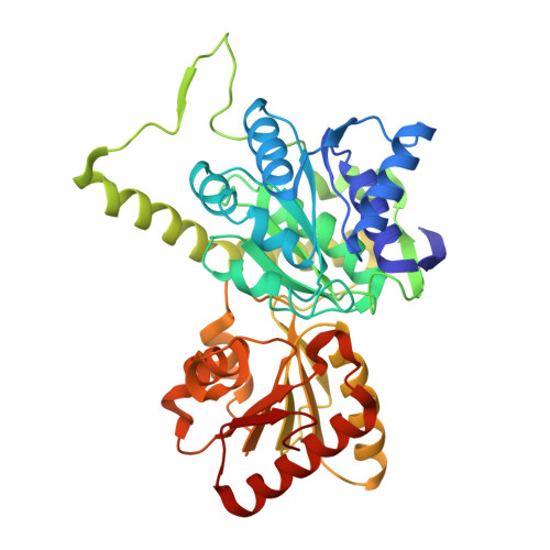
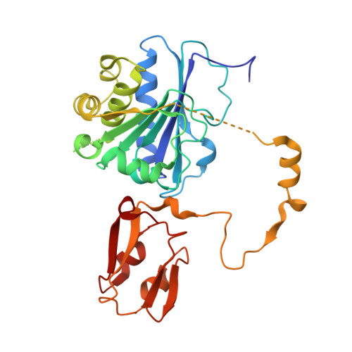
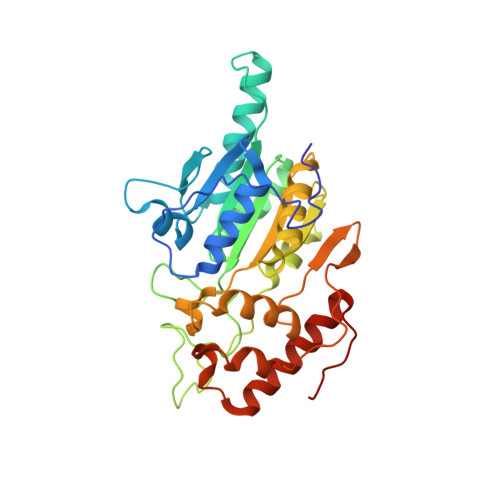
One-carbon chemistry of oxalate oxidoreductase captured by X-ray crystallography.

Gibson, M.I.Chen, P.Y.Johnson, A.C.Pierce, E.Can, M.Ragsdale, S.W.Drennan, C.L.

(2016) Proc Natl Acad Sci U S A 113: 320-325

  • DOI: https://doi.org/10.1073/pnas.1518537113
  • Primary Citation of Related Structures:  
    5EXD, 5EXE

  • PubMed Abstract: 

    Thiamine pyrophosphate (TPP)-dependent oxalate oxidoreductase (OOR) metabolizes oxalate, generating two molecules of CO2 and two low-potential electrons, thus providing both the carbon and reducing equivalents for operation of the Wood-Ljungdahl pathway of acetogenesis. Here we present structures of OOR in which two different reaction intermediate bound states have been trapped: the covalent adducts between TPP and oxalate and between TPP and CO2. These structures, along with the previously determined structure of substrate-free OOR, allow us to visualize how active site rearrangements can drive catalysis. Our results suggest that OOR operates via a bait-and-switch mechanism, attracting substrate into the active site through the presence of positively charged and polar residues, and then altering the electrostatic environment through loop and side chain movements to drive catalysis. This simple but elegant mechanism explains how oxalate, a molecule that humans and most animals cannot break down, can be used for growth by acetogenic bacteria.


  • Organizational Affiliation

    Department of Chemistry, Massachusetts Institute of Technology, Cambridge, MA 02139;


Macromolecules
Find similar proteins by:  (by identity cutoff)  |  3D Structure
Entity ID: 1
MoleculeChains Sequence LengthOrganismDetailsImage
Oxalate oxidoreductase subunit alpha
A, D, G, J
395Moorella thermoacetica ATCC 39073Mutation(s): 0 
EC: 1.2.7.10
UniProt
Find proteins for Q2RI41 (Moorella thermoacetica (strain ATCC 39073 / JCM 9320))
Explore Q2RI41 
Go to UniProtKB:  Q2RI41
Entity Groups  
Sequence Clusters30% Identity50% Identity70% Identity90% Identity95% Identity100% Identity
UniProt GroupQ2RI41
Sequence Annotations
Expand
  • Reference Sequence
Find similar proteins by:  (by identity cutoff)  |  3D Structure
Entity ID: 2
MoleculeChains Sequence LengthOrganismDetailsImage
Oxalate oxidoreductase subunit delta
B, E, H, K
315Moorella thermoacetica ATCC 39073Mutation(s): 0 
EC: 1.2.7.10
UniProt
Find proteins for Q2RI40 (Moorella thermoacetica (strain ATCC 39073 / JCM 9320))
Explore Q2RI40 
Go to UniProtKB:  Q2RI40
Entity Groups  
Sequence Clusters30% Identity50% Identity70% Identity90% Identity95% Identity100% Identity
UniProt GroupQ2RI40
Sequence Annotations
Expand
  • Reference Sequence
Find similar proteins by:  (by identity cutoff)  |  3D Structure
Entity ID: 3
MoleculeChains Sequence LengthOrganismDetailsImage
Oxalate oxidoreductase subunit beta
C, F, I, L
314Moorella thermoacetica ATCC 39073Mutation(s): 0 
EC: 1.2.7.10
UniProt
Find proteins for Q2RI42 (Moorella thermoacetica (strain ATCC 39073 / JCM 9320))
Explore Q2RI42 
Go to UniProtKB:  Q2RI42
Entity Groups  
Sequence Clusters30% Identity50% Identity70% Identity90% Identity95% Identity100% Identity
UniProt GroupQ2RI42
Sequence Annotations
Expand
  • Reference Sequence
Small Molecules
Ligands 4 Unique
IDChains Name / Formula / InChI Key2D Diagram3D Interactions
O2T
Query on O2T

Download Ideal Coordinates CCD File 
U [auth F][2-[3-[(4-azanyl-2-methyl-pyrimidin-5-yl)methyl]-4-methyl-2-[1,1,2-tris(oxidanyl)-2-oxidanylidene-ethyl]-1,3-thiazol-3-ium-5-yl]ethoxy-oxidanyl-phosphoryl] hydrogen phosphate
C14 H20 N4 O11 P2 S
DVVUFOVKVOOQQB-UHFFFAOYSA-N
TPP
Query on TPP

Download Ideal Coordinates CCD File 
EA [auth L],
P [auth C],
Z [auth I]
THIAMINE DIPHOSPHATE
C12 H19 N4 O7 P2 S
AYEKOFBPNLCAJY-UHFFFAOYSA-O
SF4
Query on SF4

Download Ideal Coordinates CCD File 
BA [auth K]
CA [auth K]
DA [auth L]
M [auth B]
N [auth B]
BA [auth K],
CA [auth K],
DA [auth L],
M [auth B],
N [auth B],
O [auth C],
R [auth E],
S [auth E],
T [auth F],
W [auth H],
X [auth H],
Y [auth I]
IRON/SULFUR CLUSTER
Fe4 S4
LJBDFODJNLIPKO-UHFFFAOYSA-N
MG
Query on MG

Download Ideal Coordinates CCD File 
AA [auth I],
FA [auth L],
Q [auth C],
V [auth F]
MAGNESIUM ION
Mg
JLVVSXFLKOJNIY-UHFFFAOYSA-N
Experimental Data & Validation

Experimental Data

  • Method: X-RAY DIFFRACTION
  • Resolution: 2.50 Å
  • R-Value Free: 0.229 
  • R-Value Work: 0.196 
  • R-Value Observed: 0.207 
  • Space Group: P 43
Unit Cell:
Length ( Å )Angle ( ˚ )
a = 138.43α = 90
b = 138.43β = 90
c = 217.069γ = 90
Software Package:
Software NamePurpose
PHENIXrefinement
HKL-2000data reduction
HKL-2000data scaling
Cootmodel building
PHENIXphasing

Structure Validation

View Full Validation Report


Ligand Structure Quality Assessment 


Entry History 

Deposition Data

Revision History  (Full details and data files)

  • Version 1.0: 2015-12-30
    Type: Initial release
  • Version 1.1: 2016-01-13
    Changes: Data collection
  • Version 1.2: 2016-01-27
    Changes: Database references
  • Version 1.3: 2023-09-27
    Changes: Data collection, Database references, Derived calculations, Refinement description