5EFQ | pdb_00005efq

Crystal structure of human Cdk13/Cyclin K in complex with ADP-aluminum fluoride


Experimental Data Snapshot

  • Method: X-RAY DIFFRACTION
  • Resolution: 2.00 Å
  • R-Value Free: 
    0.247 (Depositor) 
  • R-Value Work: 
    0.194 (Depositor) 
  • R-Value Observed: 
    0.197 (Depositor) 

Starting Model: experimental
View more details

wwPDB Validation 3D Report Full Report

Validation slider image for 5EFQ

Ligand Structure Quality Assessment 


This is version 1.4 of the entry. See complete history

Literature

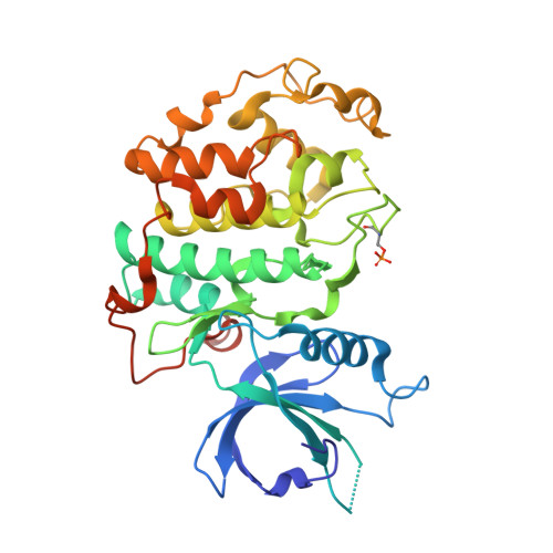
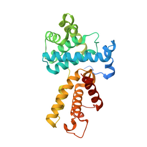
Structural and Functional Analysis of the Cdk13/Cyclin K Complex.

Greifenberg, A.K.Honig, D.Pilarova, K.Duster, R.Bartholomeeusen, K.Bosken, C.A.Anand, K.Blazek, D.Geyer, M.

(2016) Cell Rep 14: 320-331

  • DOI: https://doi.org/10.1016/j.celrep.2015.12.025
  • Primary Citation Related Structures: 
    5EFQ

  • PubMed Abstract: 

    Cyclin-dependent kinases regulate the cell cycle and transcription in higher eukaryotes. We have determined the crystal structure of the transcription kinase Cdk13 and its Cyclin K subunit at 2.0 Å resolution. Cdk13 contains a C-terminal extension helix composed of a polybasic cluster and a DCHEL motif that interacts with the bound ATP. Cdk13/CycK phosphorylates both Ser5 and Ser2 of the RNA polymerase II C-terminal domain (CTD) with a preference for Ser7 pre-phosphorylations at a C-terminal position. The peptidyl-prolyl isomerase Pin1 does not change the phosphorylation specificities of Cdk9, Cdk12, and Cdk13 but interacts with the phosphorylated CTD through its WW domain. Using recombinant proteins, we find that flavopiridol inhibits Cdk7 more potently than it does Cdk13. Gene expression changes after knockdown of Cdk13 or Cdk12 are markedly different, with enrichment of growth signaling pathways for Cdk13-dependent genes. Together, our results provide insights into the structure, function, and activity of human Cdk13/CycK.


  • Organizational Affiliation
    • Institute of Innate Immunity, Department of Structural Immunology, University of Bonn, Sigmund-Freud-Strasse 25, 53127 Bonn, Germany; Center of Advanced European Studies and Research, Group Physical Biochemistry, Ludwig-Erhard-Allee 2, 53175 Bonn, Germany.

Macromolecule Content 

  • Total Structure Weight: 145.02 kDa 
  • Atom Count: 9,353 
  • Modeled Residue Count: 1,129 
  • Deposited Residue Count: 1,230 
  • Unique protein chains: 2

Macromolecules

Find similar proteins by:|  3D Structure
Entity ID: 1
MoleculeChains  Sequence LengthOrganismDetailsImage
Cyclin-dependent kinase 13
A, C
347Homo sapiensMutation(s): 0 
Gene Names: CDK13CDC2LCDC2L5CHEDKIAA1791
EC: 2.7.11.22 (PDB Primary Data), 2.7.11.23 (PDB Primary Data)
UniProt & NIH Common Fund Data Resources
Find proteins for Q14004 (Homo sapiens)
Explore Q14004 
Go to UniProtKB:  Q14004
PHAROS:  Q14004
GTEx:  ENSG00000065883 
Entity Groups
Sequence Clusters30% Identity50% Identity70% Identity90% Identity95% Identity100% Identity
UniProt GroupQ14004
Sequence Annotations
Expand
Reference Sequence
Find similar proteins by:|  3D Structure
Entity ID: 2
MoleculeChains  Sequence LengthOrganismDetailsImage
Cyclin-K
B, D
268Homo sapiensMutation(s): 0 
Gene Names: CCNKCPR4
UniProt & NIH Common Fund Data Resources
Find proteins for O75909 (Homo sapiens)
Explore O75909 
Go to UniProtKB:  O75909
PHAROS:  O75909
GTEx:  ENSG00000090061 
Entity Groups
Sequence Clusters30% Identity50% Identity70% Identity90% Identity95% Identity100% Identity
UniProt GroupO75909
Sequence Annotations
Expand
Reference Sequence

Small Molecules

Ligands 3 Unique
IDChains Name / Formula / InChI Key2D Diagram3D Interactions
ADP

Query on ADP


Download:Ideal Coordinates CCD File
E [auth A],
I [auth C]
ADENOSINE-5'-DIPHOSPHATE
C10 H15 N5 O10 P2
XTWYTFMLZFPYCI-KQYNXXCUSA-N
AF3

Query on AF3


Download:Ideal Coordinates CCD File
F [auth A],
J [auth C]
ALUMINUM FLUORIDE
Al F3
KLZUFWVZNOTSEM-UHFFFAOYSA-K
MG

Query on MG


Download:Ideal Coordinates CCD File
G [auth A],
H [auth A],
K [auth C],
L [auth C]
MAGNESIUM ION
Mg
JLVVSXFLKOJNIY-UHFFFAOYSA-N
Modified Residues  1 Unique
IDChains TypeFormula2D DiagramParent
TPO
Query on TPO
A, C
L-PEPTIDE LINKINGC4 H10 N O6 PTHR

Experimental Data & Validation

Experimental Data

  • Method: X-RAY DIFFRACTION
  • Resolution: 2.00 Å
  • R-Value Free:  0.247 (Depositor) 
  • R-Value Work:  0.194 (Depositor) 
  • R-Value Observed: 0.197 (Depositor) 
Space Group: P 1
Unit Cell:
Length ( Å )Angle ( ˚ )
a = 50.78α = 74.08
b = 81.82β = 84.26
c = 93.14γ = 76.89
Software Package:
Software NamePurpose
PHENIXrefinement
XDSdata reduction
XDSdata scaling
PHASERphasing

Structure Validation

View Full Validation Report


Ligand Structure Quality Assessment 


Entry History 

Deposition Data

Revision History  (Full details and data files)

  • Version 1.0: 2015-12-30
    Type: Initial release
  • Version 1.1: 2016-01-20
    Changes: Database references
  • Version 1.2: 2016-01-27
    Changes: Database references
  • Version 1.3: 2024-01-10
    Changes: Data collection, Database references, Derived calculations, Refinement description
  • Version 1.4: 2024-10-23
    Changes: Structure summary