5EDI | pdb_00005edi

CRYSTAL STRUCTURE OF HUMAN PHOSPHODIESTERASE 10 IN COMPLEX WITH c2(c(n1nc(nc1c(c2)C)CCc3nc(nn3C)N4CCCC4)C)Cl, micromolar IC50=0.000279


Experimental Data Snapshot

  • Method: X-RAY DIFFRACTION
  • Resolution: 2.20 Å
  • R-Value Free: 
    0.235 (Depositor), 0.235 (DCC) 
  • R-Value Work: 
    0.186 (Depositor), 0.185 (DCC) 
  • R-Value Observed: 
    0.188 (Depositor) 

wwPDB Validation 3D Report Full Report

Validation slider image for 5EDI

Ligand Structure Quality Assessment 


This is version 1.3 of the entry. See complete history

Literature

A Real-World Perspective on Molecular Design.

Kuhn, B.Guba, W.Hert, J.Banner, D.Bissantz, C.Ceccarelli, S.Haap, W.Korner, M.Kuglstatter, A.Lerner, C.Mattei, P.Neidhart, W.Pinard, E.Rudolph, M.G.Schulz-Gasch, T.Woltering, T.Stahl, M.

(2016) J Med Chem 59: 4087-4102

  • DOI: https://doi.org/10.1021/acs.jmedchem.5b01875
  • Primary Citation Related Structures: 
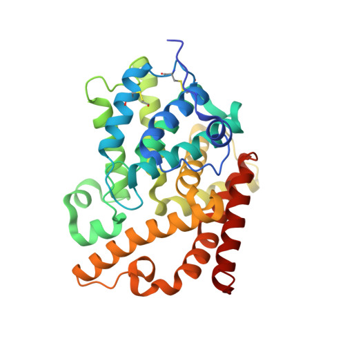
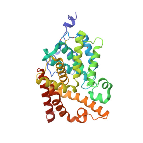
    5EDB, 5EDC, 5EDE, 5EDG, 5EDH, 5EDI, 5EZX, 5EZZ, 5F00, 5F01, 5F02, 5F03, 5I2R

  • PubMed Abstract: 

    We present a series of small molecule drug discovery case studies where computational methods were prospectively employed to impact Roche research projects, with the aim of highlighting those methods that provide real added value. Our brief accounts encompass a broad range of methods and techniques applied to a variety of enzymes and receptors. Most of these are based on judicious application of knowledge about molecular conformations and interactions: filling of lipophilic pockets to gain affinity or selectivity, addition of polar substituents, scaffold hopping, transfer of SAR, conformation analysis, and molecular overlays. A case study of sequence-driven focused screening is presented to illustrate how appropriate preprocessing of information enables effective exploitation of prior knowledge. We conclude that qualitative statements enabling chemists to focus on promising regions of chemical space are often more impactful than quantitative prediction.


  • Organizational Affiliation
    • Roche Pharmaceutical Research and Early Development, Roche Innovation Center Basel, F. Hoffmann-La Roche Ltd. , Grenzacherstrasse 124, 4070 Basel, Switzerland.

Macromolecule Content 

  • Total Structure Weight: 150.23 kDa 
  • Atom Count: 10,768 
  • Modeled Residue Count: 1,263 
  • Deposited Residue Count: 1,277 
  • Unique protein chains: 2

Macromolecules

Find similar proteins by:|  3D Structure
Entity ID: 1
MoleculeChains  Sequence LengthOrganismDetailsImage
cAMP and cAMP-inhibited cGMP 3',5'-cyclic phosphodiesterase 10A
A, C, D
319Homo sapiensMutation(s): 0 
Gene Names: PDE10A
EC: 3.1.4.17 (PDB Primary Data), 3.1.4.35 (PDB Primary Data)
UniProt & NIH Common Fund Data Resources
Find proteins for Q9Y233 (Homo sapiens)
Explore Q9Y233 
Go to UniProtKB:  Q9Y233
PHAROS:  Q9Y233
GTEx:  ENSG00000112541 
Entity Groups
Sequence Clusters30% Identity50% Identity70% Identity90% Identity95% Identity100% Identity
UniProt GroupQ9Y233
Sequence Annotations
Expand
Reference Sequence
Find similar proteins by:|  3D Structure
Entity ID: 2
MoleculeChains  Sequence LengthOrganismDetailsImage
cAMP and cAMP-inhibited cGMP 3',5'-cyclic phosphodiesterase 10A320Homo sapiensMutation(s): 0 
Gene Names: PDE10A
EC: 3.1.4.17 (PDB Primary Data), 3.1.4.35 (PDB Primary Data)
UniProt & NIH Common Fund Data Resources
Find proteins for Q9Y233 (Homo sapiens)
Explore Q9Y233 
Go to UniProtKB:  Q9Y233
PHAROS:  Q9Y233
GTEx:  ENSG00000112541 
Entity Groups
Sequence Clusters30% Identity50% Identity70% Identity90% Identity95% Identity100% Identity
UniProt GroupQ9Y233
Sequence Annotations
Expand
Reference Sequence

Small Molecules

Ligands 3 Unique
IDChains Name / Formula / InChI Key2D Diagram3D Interactions
5M9

Query on 5M9


Download:Ideal Coordinates CCD File
G [auth A],
J [auth B],
M [auth C],
P [auth D]
6-chloranyl-5,8-dimethyl-2-[2-(2-methyl-5-pyrrolidin-1-yl-1,2,4-triazol-3-yl)ethyl]-[1,2,4]triazolo[1,5-a]pyridine
C17 H22 Cl N7
NCSCZUVPQAFJGQ-UHFFFAOYSA-N
ZN

Query on ZN


Download:Ideal Coordinates CCD File
E [auth A],
H [auth B],
K [auth C],
N [auth D]
ZINC ION
Zn
PTFCDOFLOPIGGS-UHFFFAOYSA-N
MG

Query on MG


Download:Ideal Coordinates CCD File
F [auth A],
I [auth B],
L [auth C],
O [auth D]
MAGNESIUM ION
Mg
JLVVSXFLKOJNIY-UHFFFAOYSA-N
Modified Residues  1 Unique
IDChains TypeFormula2D DiagramParent
CME
Query on CME
A, C, D
L-PEPTIDE LINKINGC5 H11 N O3 S2CYS

Experimental Data & Validation

Experimental Data

  • Method: X-RAY DIFFRACTION
  • Resolution: 2.20 Å
  • R-Value Free:  0.235 (Depositor), 0.235 (DCC) 
  • R-Value Work:  0.186 (Depositor), 0.185 (DCC) 
  • R-Value Observed: 0.188 (Depositor) 
Space Group: H 3
Unit Cell:
Length ( Å )Angle ( ˚ )
a = 135.231α = 90
b = 135.231β = 90
c = 235.395γ = 120
Software Package:
Software NamePurpose
XSCALEdata scaling
PDB_EXTRACTdata extraction
REFMACrefinement
XDSdata reduction
REFMACphasing

Structure Validation

View Full Validation Report


Ligand Structure Quality Assessment 


Entry History 

Deposition Data

Revision History  (Full details and data files)

  • Version 1.0: 2016-03-09
    Type: Initial release
  • Version 1.1: 2016-05-25
    Changes: Database references
  • Version 1.2: 2024-10-09
    Changes: Data collection, Database references, Derived calculations, Structure summary
  • Version 1.3: 2024-10-16
    Changes: Structure summary