5E11

Second PDZ domain of Ligand of Numb protein X 2 by Laue crystallography (no electric field)


Experimental Data Snapshot

  • Method: X-RAY DIFFRACTION
  • Resolution: 1.80 Å
  • R-Value Free: 0.148 
  • R-Value Work: 0.132 
  • R-Value Observed: 0.133 

wwPDB Validation   3D Report Full Report


This is version 1.3 of the entry. See complete history


Literature

Electric-field-stimulated protein mechanics.

Hekstra, D.R.White, K.I.Socolich, M.A.Henning, R.W.Srajer, V.Ranganathan, R.

(2016) Nature 540: 400-405

  • DOI: https://doi.org/10.1038/nature20571
  • Primary Citation of Related Structures:  
    5E11, 5E1Y, 5E21, 5E22

  • PubMed Abstract: 

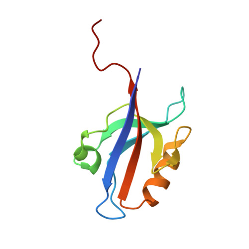
    The internal mechanics of proteins-the coordinated motions of amino acids and the pattern of forces constraining these motions-connects protein structure to function. Here we describe a new method combining the application of strong electric field pulses to protein crystals with time-resolved X-ray crystallography to observe conformational changes in spatial and temporal detail. Using a human PDZ domain (LNX2 PDZ2 ) as a model system, we show that protein crystals tolerate electric field pulses strong enough to drive concerted motions on the sub-microsecond timescale. The induced motions are subtle, involve diverse physical mechanisms, and occur throughout the protein structure. The global pattern of electric-field-induced motions is consistent with both local and allosteric conformational changes naturally induced by ligand binding, including at conserved functional sites in the PDZ domain family. This work lays the foundation for comprehensive experimental study of the mechanical basis of protein function.


  • Organizational Affiliation

    Green Center for Systems Biology, UT Southwestern Medical Center, 6001 Forest Park Road, Dallas, Texas 75390, USA.


Macromolecules
Find similar proteins by:  (by identity cutoff)  |  3D Structure
Entity ID: 1
MoleculeChains Sequence LengthOrganismDetailsImage
Ligand of Numb protein X 295Homo sapiensMutation(s): 1 
Gene Names: LNX2PDZRN1
UniProt & NIH Common Fund Data Resources
Find proteins for Q8N448 (Homo sapiens)
Explore Q8N448 
Go to UniProtKB:  Q8N448
PHAROS:  Q8N448
GTEx:  ENSG00000139517 
Entity Groups  
Sequence Clusters30% Identity50% Identity70% Identity90% Identity95% Identity100% Identity
UniProt GroupQ8N448
Sequence Annotations
Expand
  • Reference Sequence
Experimental Data & Validation

Experimental Data

  • Method: X-RAY DIFFRACTION
  • Resolution: 1.80 Å
  • R-Value Free: 0.148 
  • R-Value Work: 0.132 
  • R-Value Observed: 0.133 
  • Space Group: C 1 2 1
Unit Cell:
Length ( Å )Angle ( ˚ )
a = 65.3α = 90
b = 39.45β = 117.54
c = 39.01γ = 90
Software Package:
Software NamePurpose
PHENIXrefinement
Precognitiondata reduction
Epinormdata reduction
PHENIXphasing

Structure Validation

View Full Validation Report


Entry History & Funding Information

Deposition Data


Funding OrganizationLocationGrant Number
Robert A. Welch FoundationI-1366

Revision History  (Full details and data files)

  • Version 1.0: 2016-12-07
    Type: Initial release
  • Version 1.1: 2017-01-11
    Changes: Database references
  • Version 1.2: 2017-11-22
    Changes: Refinement description
  • Version 1.3: 2024-03-06
    Changes: Data collection, Database references