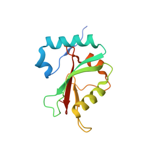
5DPT

Crystal structure of PLEKHM1 LIR-fused human GABARAPL1_2-117


Experimental Data Snapshot

  • Method: X-RAY DIFFRACTION
  • Resolution: 2.90 Å
  • R-Value Free: 0.260 
  • R-Value Work: 0.214 
  • R-Value Observed: 0.216 

wwPDB Validation   3D Report Full Report


This is version 1.2 of the entry. See complete history


Literature

Structural and functional analysis of the GABARAP interaction motif (GIM).

Rogov, V.V.Stolz, A.Ravichandran, A.C.Rios-Szwed, D.O.Suzuki, H.Kniss, A.Lohr, F.Wakatsuki, S.Dotsch, V.Dikic, I.Dobson, R.C.McEwan, D.G.

(2017) EMBO Rep 18: 1382-1396

  • DOI: https://doi.org/10.15252/embr.201643587
  • Primary Citation of Related Structures:  
    5DPR, 5DPS, 5DPT, 5DPW

  • PubMed Abstract: 

    Through the canonical LC3 interaction motif (LIR), [W/F/Y]-X 1 -X 2 -[I/L/V], protein complexes are recruited to autophagosomes to perform their functions as either autophagy adaptors or receptors. How these adaptors/receptors selectively interact with either LC3 or GABARAP families remains unclear. Herein, we determine the range of selectivity of 30 known core LIR motifs towards individual LC3s and GABARAPs. From these, we define a G ABARAP I nteraction M otif (GIM) sequence ([W/F]-[V/I]-X 2 -V) that the adaptor protein PLEKHM1 tightly conforms to. Using biophysical and structural approaches, we show that the PLEKHM1-LIR is indeed 11-fold more specific for GABARAP than LC3B. Selective mutation of the X 1 and X 2 positions either completely abolished the interaction with all LC3 and GABARAPs or increased PLEKHM1-GIM selectivity 20-fold towards LC3B. Finally, we show that conversion of p62/SQSTM1, FUNDC1 and FIP200 LIRs into our newly defined GIM, by introducing two valine residues, enhances their interaction with endogenous GABARAP over LC3B. The identification of a GABARAP-specific interaction motif will aid the identification and characterization of the expanding array of autophagy receptor and adaptor proteins and their in vivo functions.


  • Organizational Affiliation

    Institute of Biophysical Chemistry and Center for Biomolecular Magnetic Resonance, Goethe University, Frankfurt am Main, Germany.


Macromolecules
Find similar proteins by:  (by identity cutoff)  |  3D Structure
Entity ID: 1
MoleculeChains Sequence LengthOrganismDetailsImage
Pleckstrin homology domain-containing family M member 1, Gamma-aminobutyric acid receptor-associated protein-like 1,Gamma-aminobutyric acid receptor-associated protein-like 1
A, B
132Homo sapiensMutation(s): 0 
Gene Names: PLEKHM1GABARAPL1
UniProt & NIH Common Fund Data Resources
Find proteins for Q9H0R8 (Homo sapiens)
Explore Q9H0R8 
Go to UniProtKB:  Q9H0R8
PHAROS:  Q9H0R8
GTEx:  ENSG00000139112 
Find proteins for Q9Y4G2 (Homo sapiens)
Explore Q9Y4G2 
Go to UniProtKB:  Q9Y4G2
PHAROS:  Q9Y4G2
GTEx:  ENSG00000225190 
Entity Groups  
Sequence Clusters30% Identity50% Identity70% Identity90% Identity95% Identity100% Identity
UniProt GroupsQ9Y4G2Q9H0R8
Sequence Annotations
Expand
  • Reference Sequence
Small Molecules
Experimental Data & Validation

Experimental Data

  • Method: X-RAY DIFFRACTION
  • Resolution: 2.90 Å
  • R-Value Free: 0.260 
  • R-Value Work: 0.214 
  • R-Value Observed: 0.216 
  • Space Group: P 32 2 1
Unit Cell:
Length ( Å )Angle ( ˚ )
a = 84.791α = 90
b = 84.791β = 90
c = 105.44γ = 120
Software Package:
Software NamePurpose
PHENIXrefinement
XDSdata reduction
Aimlessdata scaling
MOLREPphasing

Structure Validation

View Full Validation Report


Entry History 

Deposition Data

Revision History  (Full details and data files)

  • Version 1.0: 2016-09-28
    Type: Initial release
  • Version 1.1: 2018-06-20
    Changes: Data collection, Database references, Derived calculations
  • Version 1.2: 2024-03-06
    Changes: Data collection, Database references