5DMP

Structure of the Archaeal NHEJ Phosphoesterase from Methanocella paludicola.


Experimental Data Snapshot

  • Method: X-RAY DIFFRACTION
  • Resolution: 1.79 Å
  • R-Value Free: 0.204 
  • R-Value Work: 0.174 
  • R-Value Observed: 0.176 

wwPDB Validation   3D Report Full Report


This is version 1.2 of the entry. See complete history


Literature

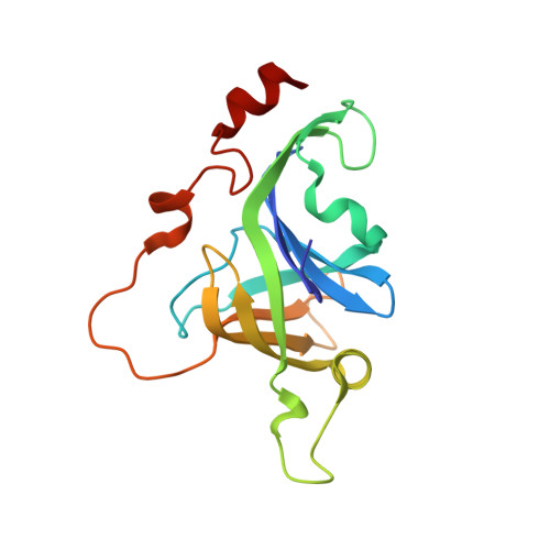
Molecular basis for DNA strand displacement by NHEJ repair polymerases.

Bartlett, E.J.Brissett, N.C.Plocinski, P.Carlberg, T.Doherty, A.J.

(2016) Nucleic Acids Res 44: 2173-2186

  • DOI: https://doi.org/10.1093/nar/gkv965
  • Primary Citation of Related Structures:  
    5DMP, 5DMU

  • PubMed Abstract: 

    The non-homologous end-joining (NHEJ) pathway repairs DNA double-strand breaks (DSBs) in all domains of life. Archaea and bacteria utilize a conserved set of multifunctional proteins in a pathway termed Archaeo-Prokaryotic (AP) NHEJ that facilitates DSB repair. Archaeal NHEJ polymerases (Pol) are capable of strand displacement synthesis, whilst filling DNA gaps or partially annealed DNA ends, which can give rise to unligatable intermediates. However, an associated NHEJ phosphoesterase (PE) resects these products to ensure that efficient ligation occurs. Here, we describe the crystal structures of these archaeal (Methanocella paludicola) NHEJ nuclease and polymerase enzymes, demonstrating their strict structural conservation with their bacterial NHEJ counterparts. Structural analysis, in conjunction with biochemical studies, has uncovered the molecular basis for DNA strand displacement synthesis in AP-NHEJ, revealing the mechanisms that enable Pol and PE to displace annealed bases to facilitate their respective roles in DSB repair.


  • Organizational Affiliation

    Genome Damage and Stability Centre, School of Life Sciences, University of Sussex, Brighton, BN1 9RQ, UK.


Macromolecules
Find similar proteins by:  (by identity cutoff)  |  3D Structure
Entity ID: 1
MoleculeChains Sequence LengthOrganismDetailsImage
Uncharacterized protein169Methanocella paludicolaMutation(s): 0 
Gene Names: MCP_2127
UniProt
Find proteins for D1Z0H7 (Methanocella paludicola (strain DSM 17711 / JCM 13418 / NBRC 101707 / SANAE))
Explore D1Z0H7 
Go to UniProtKB:  D1Z0H7
Entity Groups  
Sequence Clusters30% Identity50% Identity70% Identity90% Identity95% Identity100% Identity
UniProt GroupD1Z0H7
Sequence Annotations
Expand
  • Reference Sequence
Small Molecules
Ligands 3 Unique
IDChains Name / Formula / InChI Key2D Diagram3D Interactions
VN4
Query on VN4

Download Ideal Coordinates CCD File 
J [auth A]oxido(dioxo)vanadium
O3 V
ALTWGIIQPLQAAM-UHFFFAOYSA-N
EDO
Query on EDO

Download Ideal Coordinates CCD File 
B [auth A]
C [auth A]
D [auth A]
E [auth A]
F [auth A]
B [auth A],
C [auth A],
D [auth A],
E [auth A],
F [auth A],
G [auth A],
H [auth A]
1,2-ETHANEDIOL
C2 H6 O2
LYCAIKOWRPUZTN-UHFFFAOYSA-N
MG
Query on MG

Download Ideal Coordinates CCD File 
I [auth A]MAGNESIUM ION
Mg
JLVVSXFLKOJNIY-UHFFFAOYSA-N
Experimental Data & Validation

Experimental Data

  • Method: X-RAY DIFFRACTION
  • Resolution: 1.79 Å
  • R-Value Free: 0.204 
  • R-Value Work: 0.174 
  • R-Value Observed: 0.176 
  • Space Group: P 31 2 1
Unit Cell:
Length ( Å )Angle ( ˚ )
a = 57.07α = 90
b = 57.07β = 90
c = 105.041γ = 120
Software Package:
Software NamePurpose
CrystalCleardata collection
SCALAdata scaling
PHASERphasing
REFMACrefinement
PDB_EXTRACTdata extraction
MOSFLMdata reduction
Cootmodel building

Structure Validation

View Full Validation Report


Entry History & Funding Information

Deposition Data


Funding OrganizationLocationGrant Number
Biotechnology and Biological Sciences Research CouncilUnited Kingdom(BB/J018643/1)

Revision History  (Full details and data files)

  • Version 1.0: 2015-10-07
    Type: Initial release
  • Version 1.1: 2016-03-30
    Changes: Database references
  • Version 1.2: 2024-01-10
    Changes: Data collection, Database references, Refinement description