5DF9

CRYSTAL STRUCTURE OF PENICILLIN-BINDING PROTEIN 3 IN COMPLEX WITH DEACYLATED PRODUCT OF CEFOPERAZONE


Experimental Data Snapshot

  • Method: X-RAY DIFFRACTION
  • Resolution: 2.70 Å
  • R-Value Free: 0.249 
  • R-Value Work: 0.202 
  • R-Value Observed: 0.205 

wwPDB Validation   3D Report Full Report


Ligand Structure Quality Assessment 


This is version 1.2 of the entry. See complete history


Literature

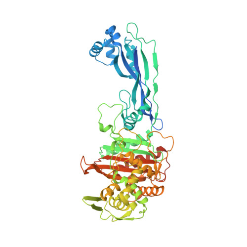
Crystal structures of penicillin-binding protein 3 in complexes with azlocillin and cefoperazone in both acylated and deacylated forms.

Ren, J.Nettleship, J.E.Males, A.Stuart, D.I.Owens, R.J.

(2016) FEBS Lett 590: 288-297

  • DOI: https://doi.org/10.1002/1873-3468.12054
  • Primary Citation of Related Structures:  
    5DF7, 5DF8, 5DF9

  • PubMed Abstract: 

    Penicillin-binding protein 3 (PBP3) from Pseudomonas aeruginosa is the molecular target of β-lactam-based antibiotics. Structures of PBP3 in complexes with azlocillin and cefoperazone, which are in clinical use for the treatment of pseudomonad infections, have been determined to 2.0 Å resolution. Together with data from other complexes, these structures identify a common set of residues involved in the binding of β-lactams to PBP3. Comparison of wild-type and an active site mutant (S294A) showed that increased thermal stability of PBP3 following azlocillin binding was entirely due to covalent binding to S294, whereas cefoperazone binding produces some increase in stability without the covalent link. Consistent with this, a third crystal structure was determined in which the hydrolysis product of cefoperazone was noncovalently bound in the active site of PBP3. This is the first structure of a complex between a penicillin-binding protein and cephalosporic acid and may be important in the design of new noncovalent PBP3 inhibitors.


  • Organizational Affiliation

    Division of Structural Biology, Henry Wellcome Building for Genomic Medicine, University of Oxford, Oxford, UK.


Macromolecules
Find similar proteins by:  (by identity cutoff)  |  3D Structure
Entity ID: 1
MoleculeChains Sequence LengthOrganismDetailsImage
Cell division protein564Pseudomonas aeruginosaMutation(s): 0 
Gene Names: pbpBftsI_2ERS445055_04698PAE221_03076YQ19_27590
EC: 2.4.1.129
UniProt
Find proteins for Q51504 (Pseudomonas aeruginosa)
Explore Q51504 
Go to UniProtKB:  Q51504
Entity Groups  
Sequence Clusters30% Identity50% Identity70% Identity90% Identity95% Identity100% Identity
UniProt GroupQ51504
Sequence Annotations
Expand
  • Reference Sequence
Small Molecules
Ligands 3 Unique
IDChains Name / Formula / InChI Key2D Diagram3D Interactions
59J
Query on 59J

Download Ideal Coordinates CCD File 
D [auth A](2R,5R)-2-[(R)-carboxy{[(2R)-2-{[(4-ethyl-2,3-dioxopiperazin-1-yl)carbonyl]amino}-2-(4-hydroxyphenyl)acetyl]amino}methyl]-5-methyl-5,6-dihydro-2H-1,3-thiazine-4-carboxylic acid
C23 H27 N5 O9 S
YNTRKCUUJNUMAH-QOQPWIFLSA-N
SO4
Query on SO4

Download Ideal Coordinates CCD File 
B [auth A],
C [auth A]
SULFATE ION
O4 S
QAOWNCQODCNURD-UHFFFAOYSA-L
GOL
Query on GOL

Download Ideal Coordinates CCD File 
E [auth A]GLYCEROL
C3 H8 O3
PEDCQBHIVMGVHV-UHFFFAOYSA-N
Experimental Data & Validation

Experimental Data

  • Method: X-RAY DIFFRACTION
  • Resolution: 2.70 Å
  • R-Value Free: 0.249 
  • R-Value Work: 0.202 
  • R-Value Observed: 0.205 
  • Space Group: C 1 2 1
Unit Cell:
Length ( Å )Angle ( ˚ )
a = 176.884α = 90
b = 41.264β = 117.42
c = 87.786γ = 90
Software Package:
Software NamePurpose
REFMACrefinement
HKL-2000data reduction
HKL-2000data scaling
MOLREPphasing

Structure Validation

View Full Validation Report


Ligand Structure Quality Assessment 


Entry History 

Deposition Data

Revision History  (Full details and data files)

  • Version 1.0: 2016-01-13
    Type: Initial release
  • Version 1.1: 2016-02-10
    Changes: Database references
  • Version 1.2: 2024-01-10
    Changes: Data collection, Database references, Refinement description