5DEG | pdb_00005deg

Crystal structure of B*27:06 bound to the pVIPR peptide


Experimental Data Snapshot

  • Method: X-RAY DIFFRACTION
  • Resolution: 1.83 Å
  • R-Value Free: 
    0.193 (Depositor), 0.195 (DCC) 
  • R-Value Work: 
    0.171 (Depositor), 0.174 (DCC) 
  • R-Value Observed: 
    0.172 (Depositor) 

Starting Model: experimental
View more details

wwPDB Validation 3D Report Full Report

Validation slider image for 5DEG

This is version 1.5 of the entry. See complete history

Literature

Increased Conformational Flexibility of HLA-B*27 Subtypes Associated With Ankylosing Spondylitis.

Loll, B.Fabian, H.Huser, H.Hee, C.S.Ziegler, A.Uchanska-Ziegler, B.Ziegler, A.

(2016) Arthritis Rheumatol 68: 1172-1182

  • DOI: https://doi.org/10.1002/art.39567
  • Primary Citation Related Structures: 
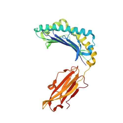
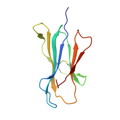
    5DEF, 5DEG

  • PubMed Abstract: 

    Dissimilarities in antigen processing and presentation are known to contribute to the differential association of HLA-B*27 subtypes with the inflammatory rheumatic disease ankylosing spondylitis (AS). In support of this notion, previous x-ray crystallographic data showed that peptides can be displayed by almost identical HLA-B*27 molecules in a subtype-dependent manner, allowing cytotoxic T lymphocytes to distinguish between these subtypes. For example, a human self-peptide derived from vasoactive intestinal peptide receptor type 1 (pVIPR; sequence RRKWRRWHL) is displayed in a single conformation by B*27:09 (which is not associated with AS), while B*27:05 (which is associated with AS) presents the peptide in a dual binding mode. In addition, differences in conformational flexibility between these subtypes might affect their stability or antigen presentation capability. This study was undertaken to investigate B*27:04 and B*27:06, another pair of minimally distinct HLA-B*27 subtypes, to assess whether dual peptide conformations or structural dynamics play a role in the initiation of AS. Using x-ray crystallography, we determined the structures of the pVIPR-B*27:04 and pVIPR-B*27:06 complexes and used isotope-edited infrared (IR) spectroscopy to probe the dynamics of these HLA-B*27 subtypes. As opposed to B*27:05 and B*27:09, B*27:04 (which is associated with AS) displays pVIPR conventionally and B*27:06 (which is not associated with AS) presents the peptide in a dual conformation. Comparison of the 4 HLA-B*27 subtypes using IR spectroscopy revealed that B*27:04 and B*27:05 possess elevated molecular dynamics compared to the nonassociated subtypes B*27:06 and B*27:09. Our results demonstrate that an increase in conformational flexibility characterizes the disease-associated subtypes B*27:04 and B*27:05.


  • Organizational Affiliation
    • Freie Universität Berlin, Berlin, Germany.

Macromolecule Content 

  • Total Structure Weight: 45.4 kDa 
  • Atom Count: 3,865 
  • Modeled Residue Count: 385 
  • Deposited Residue Count: 385 
  • Unique protein chains: 3

Macromolecules

Find similar proteins by:|  3D Structure
Entity ID: 1
MoleculeChains  Sequence LengthOrganismDetailsImage
MHC class I antigen276Homo sapiensMutation(s): 0 
Gene Names: HLA-B
UniProt & NIH Common Fund Data Resources
Find proteins for P01889 (Homo sapiens)
Explore P01889 
Go to UniProtKB:  P01889
PHAROS:  P01889
GTEx:  ENSG00000234745 
Entity Groups
Sequence Clusters30% Identity50% Identity70% Identity90% Identity95% Identity100% Identity
UniProt GroupP01889
Sequence Annotations
Expand
Reference Sequence
Find similar proteins by:|  3D Structure
Entity ID: 2
MoleculeChains  Sequence LengthOrganismDetailsImage
Beta-2-microglobulin100Homo sapiensMutation(s): 0 
Gene Names: B2MCDABP0092HDCMA22P
UniProt & NIH Common Fund Data Resources
Find proteins for P61769 (Homo sapiens)
Explore P61769 
Go to UniProtKB:  P61769
PHAROS:  P61769
GTEx:  ENSG00000166710 
Entity Groups
Sequence Clusters30% Identity50% Identity70% Identity90% Identity95% Identity100% Identity
UniProt GroupP61769
Sequence Annotations
Expand
Reference Sequence
Find similar proteins by:  Sequence   |   3D Structure  
Entity ID: 3
MoleculeChains  Sequence LengthOrganismDetailsImage
Peptide derived of VASOACTIVE INTESTINAL POLYPEPTIDE RECEPTOR 1 (pVIPR)9Homo sapiensMutation(s): 0 
UniProt & NIH Common Fund Data Resources
Find proteins for P32241 (Homo sapiens)
Explore P32241 
Go to UniProtKB:  P32241
PHAROS:  P32241
GTEx:  ENSG00000114812 
Entity Groups
UniProt GroupP32241
Sequence Annotations
Expand
Reference Sequence

Experimental Data & Validation

Experimental Data

  • Method: X-RAY DIFFRACTION
  • Resolution: 1.83 Å
  • R-Value Free:  0.193 (Depositor), 0.195 (DCC) 
  • R-Value Work:  0.171 (Depositor), 0.174 (DCC) 
  • R-Value Observed: 0.172 (Depositor) 
Space Group: P 1 21 1
Unit Cell:
Length ( Å )Angle ( ˚ )
a = 50.768α = 90
b = 81.684β = 107.53
c = 64.433γ = 90
Software Package:
Software NamePurpose
REFMACrefinement
DENZOdata reduction
SCALEPACKdata scaling
PHASERphasing

Structure Validation

View Full Validation Report


Entry History 

& Funding Information

Deposition Data


Funding OrganizationLocationGrant Number
German Research FoundationGermanySFB 449
German Research FoundationGermanyUC 8/1-3

Revision History  (Full details and data files)

  • Version 1.0: 2015-11-18
    Type: Initial release
  • Version 1.1: 2016-01-20
    Changes: Database references
  • Version 1.2: 2016-01-27
    Changes: Database references
  • Version 1.3: 2019-12-18
    Changes: Author supporting evidence, Database references
  • Version 1.4: 2024-01-10
    Changes: Data collection, Database references, Refinement description
  • Version 1.5: 2024-10-23
    Changes: Structure summary