5D2D

Crystal structure of human 14-3-3 zeta in complex with CFTR R-domain peptide pS753-pS768


Experimental Data Snapshot

  • Method: X-RAY DIFFRACTION
  • Resolution: 2.10 Å
  • R-Value Free: 0.220 
  • R-Value Work: 0.193 
  • R-Value Observed: 0.195 

wwPDB Validation   3D Report Full Report


This is version 2.0 of the entry. See complete history


Literature

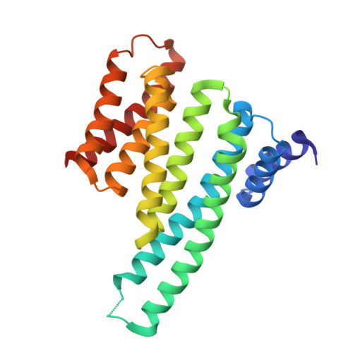
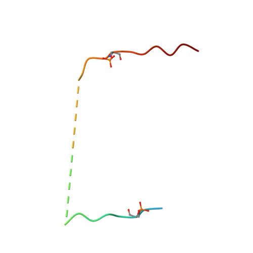
Characterization and small-molecule stabilization of the multisite tandem binding between 14-3-3 and the R domain of CFTR.

Stevers, L.M.Lam, C.V.Leysen, S.F.Meijer, F.A.van Scheppingen, D.S.de Vries, R.M.Carlile, G.W.Milroy, L.G.Thomas, D.Y.Brunsveld, L.Ottmann, C.

(2016) Proc Natl Acad Sci U S A 113: E1152-E1161

  • DOI: https://doi.org/10.1073/pnas.1516631113
  • Primary Citation of Related Structures:  
    5D2D, 5D3E, 5D3F

  • PubMed Abstract: 

    Cystic fibrosis is a fatal genetic disease, most frequently caused by the retention of the CFTR (cystic fibrosis transmembrane conductance regulator) mutant protein in the endoplasmic reticulum (ER). The binding of the 14-3-3 protein to the CFTR regulatory (R) domain has been found to enhance CFTR trafficking to the plasma membrane. To define the mechanism of action of this protein-protein interaction, we have examined the interaction in vitro. The disordered multiphosphorylated R domain contains nine different 14-3-3 binding motifs. Furthermore, the 14-3-3 protein forms a dimer containing two amphipathic grooves that can potentially bind these phosphorylated motifs. This results in a number of possible binding mechanisms between these two proteins. Using multiple biochemical assays and crystal structures, we show that the interaction between them is governed by two binding sites: The key binding site of CFTR (pS768) occupies one groove of the 14-3-3 dimer, and a weaker, secondary binding site occupies the other binding groove. We show that fusicoccin-A, a natural-product tool compound used in studies of 14-3-3 biology, can stabilize the interaction between 14-3-3 and CFTR by selectively interacting with a secondary binding motif of CFTR (pS753). The stabilization of this interaction stimulates the trafficking of mutant CFTR to the plasma membrane. This definition of the druggability of the 14-3-3-CFTR interface might offer an approach for cystic fibrosis therapeutics.


  • Organizational Affiliation

    Laboratory of Chemical Biology, Department of Biomedical Engineering, Eindhoven University of Technology, 5600 MB Eindhoven, The Netherlands; Institute for Complex Molecular Systems, Eindhoven University of Technology, 5600 MB Eindhoven, The Netherlands;


Macromolecules
Find similar proteins by:  (by identity cutoff)  |  3D Structure
Entity ID: 1
MoleculeChains Sequence LengthOrganismDetailsImage
14-3-3 protein zeta/delta
A, B
230Homo sapiensMutation(s): 0 
Gene Names: YWHAZ
UniProt & NIH Common Fund Data Resources
Find proteins for P63104 (Homo sapiens)
Explore P63104 
Go to UniProtKB:  P63104
PHAROS:  P63104
GTEx:  ENSG00000164924 
Entity Groups  
Sequence Clusters30% Identity50% Identity70% Identity90% Identity95% Identity100% Identity
UniProt GroupP63104
Sequence Annotations
Expand
  • Reference Sequence
Find similar proteins by:  (by identity cutoff)  |  3D Structure
Entity ID: 2
MoleculeChains Sequence LengthOrganismDetailsImage
Cystic fibrosis transmembrane conductance regulator28Homo sapiensMutation(s): 0 
EC: 3.6.3.49
UniProt & NIH Common Fund Data Resources
Find proteins for P13569 (Homo sapiens)
Explore P13569 
Go to UniProtKB:  P13569
PHAROS:  P13569
GTEx:  ENSG00000001626 
Entity Groups  
Sequence Clusters30% Identity50% Identity70% Identity90% Identity95% Identity100% Identity
UniProt GroupP13569
Sequence Annotations
Expand
  • Reference Sequence
Oligosaccharides

Help

Entity ID: 3
MoleculeChains Length2D Diagram Glycosylation3D Interactions
beta-D-fructofuranose-(2-1)-alpha-D-glucopyranose
D, E
2N/A
Glycosylation Resources
GlyTouCan:  G05551OP
GlyCosmos:  G05551OP
Biologically Interesting Molecules (External Reference) 1 Unique
Experimental Data & Validation

Experimental Data

  • Method: X-RAY DIFFRACTION
  • Resolution: 2.10 Å
  • R-Value Free: 0.220 
  • R-Value Work: 0.193 
  • R-Value Observed: 0.195 
  • Space Group: P 41 21 2
Unit Cell:
Length ( Å )Angle ( ˚ )
a = 112.862α = 90
b = 112.862β = 90
c = 158.192γ = 90
Software Package:
Software NamePurpose
PHENIXrefinement
XDSdata reduction
Aimlessdata scaling

Structure Validation

View Full Validation Report


Entry History & Funding Information

Deposition Data


Funding OrganizationLocationGrant Number
NWONetherlands--

Revision History  (Full details and data files)

  • Version 1.0: 2016-03-16
    Type: Initial release
  • Version 2.0: 2020-07-29
    Type: Remediation
    Reason: Carbohydrate remediation
    Changes: Atomic model, Data collection, Derived calculations, Non-polymer description, Structure summary