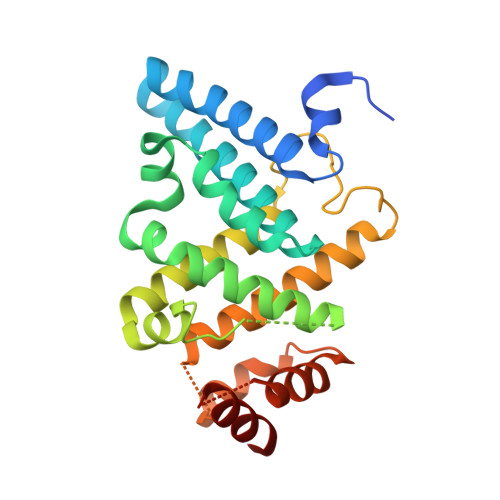
5CV1

C. elegans PGL-1 Dimerization Domain


Experimental Data Snapshot

  • Method: X-RAY DIFFRACTION
  • Resolution: 3.60 Å
  • R-Value Free: 0.321 
  • R-Value Work: 0.299 
  • R-Value Observed: 0.301 

wwPDB Validation   3D Report Full Report


This is version 1.4 of the entry. See complete history


Literature

PGL germ granule assembly protein is a base-specific, single-stranded RNase.

Aoki, S.T.Kershner, A.M.Bingman, C.A.Wickens, M.Kimble, J.

(2016) Proc Natl Acad Sci U S A 113: 1279-1284

  • DOI: https://doi.org/10.1073/pnas.1524400113
  • Primary Citation of Related Structures:  
    5COW, 5CV1, 5CV3

  • PubMed Abstract: 

    Cellular RNA-protein (RNP) granules are ubiquitous and have fundamental roles in biology and RNA metabolism, but the molecular basis of their structure, assembly, and function is poorly understood. Using nematode "P-granules" as a paradigm, we focus on the PGL granule scaffold protein to gain molecular insights into RNP granule structure and assembly. We first identify a PGL dimerization domain (DD) and determine its crystal structure. PGL-1 DD has a novel 13 α-helix fold that creates a positively charged channel as a homodimer. We investigate its capacity to bind RNA and discover unexpectedly that PGL-1 DD is a guanosine-specific, single-stranded endonuclease. Discovery of the PGL homodimer, together with previous results, suggests a model in which the PGL DD dimer forms a fundamental building block for P-granule assembly. Discovery of the PGL RNase activity expands the role of RNP granule assembly proteins to include enzymatic activity in addition to their job as structural scaffolds.


  • Organizational Affiliation

    Department of Biochemistry, University of Wisconsin-Madison, Madison, WI 53706;


Macromolecules
Find similar proteins by:  (by identity cutoff)  |  3D Structure
Entity ID: 1
MoleculeChains Sequence LengthOrganismDetailsImage
P granule abnormality protein 1243Caenorhabditis elegansMutation(s): 0 
Gene Names: pgl-1ZK381.4
UniProt
Find proteins for Q9TZQ3 (Caenorhabditis elegans)
Explore Q9TZQ3 
Go to UniProtKB:  Q9TZQ3
Entity Groups  
Sequence Clusters30% Identity50% Identity70% Identity90% Identity95% Identity100% Identity
UniProt GroupQ9TZQ3
Sequence Annotations
Expand
  • Reference Sequence
Experimental Data & Validation

Experimental Data

  • Method: X-RAY DIFFRACTION
  • Resolution: 3.60 Å
  • R-Value Free: 0.321 
  • R-Value Work: 0.299 
  • R-Value Observed: 0.301 
  • Space Group: P 61 2 2
Unit Cell:
Length ( Å )Angle ( ˚ )
a = 59.72α = 90
b = 59.72β = 90
c = 227.24γ = 120
Software Package:
Software NamePurpose
PHENIXrefinement
XSCALEdata scaling
PHASERphasing
RESOLVEmodel building
PDB_EXTRACTdata extraction
Cootmodel building

Structure Validation

View Full Validation Report


Entry History & Funding Information

Deposition Data


Funding OrganizationLocationGrant Number
Howard Hughes Medical Institute (HHMI)United States--
National Institutes of Health/Eunice Kennedy Shriver National Institute of Child Health & Human Development (NIH/NICHD)United States5F32HD071692

Revision History  (Full details and data files)

  • Version 1.0: 2016-02-03
    Type: Initial release
  • Version 1.1: 2016-02-10
    Changes: Database references
  • Version 1.2: 2017-09-06
    Changes: Author supporting evidence, Database references, Derived calculations
  • Version 1.3: 2019-11-20
    Changes: Author supporting evidence
  • Version 1.4: 2023-09-27
    Changes: Data collection, Database references, Refinement description