5CAH

Crystal structure of Mycobacterium tuberculosis malate synthase in complex with 6H-thieno[2,3-b]pyrrole-5-carboxylic acid


Experimental Data Snapshot

  • Method: X-RAY DIFFRACTION
  • Resolution: 2.30 Å
  • R-Value Free: 0.290 
  • R-Value Work: 0.202 
  • R-Value Observed: 0.206 

wwPDB Validation   3D Report Full Report


Ligand Structure Quality Assessment 


This is version 1.4 of the entry. See complete history


Literature

Mycobacterium tuberculosis Malate Synthase Structures with Fragments Reveal a Portal for Substrate/Product Exchange.

Huang, H.L.Krieger, I.V.Parai, M.K.Gawandi, V.B.Sacchettini, J.C.

(2016) J Biol Chem 291: 27421-27432

  • DOI: https://doi.org/10.1074/jbc.M116.750877
  • Primary Citation of Related Structures:  
    5C7V, 5C9R, 5C9U, 5C9W, 5C9X, 5CAH, 5CAK, 5CBB, 5CBI, 5CBJ, 5CCZ, 5CEW, 5CJM, 5CJN, 5DRC, 5DRI, 5DX7, 5E9X, 5ECV, 5H8M, 5H8P, 5H8U, 5T8G

  • PubMed Abstract: 

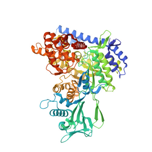
    Fragment screening and high throughput screening are complementary approaches that combine with structural biology to explore the binding capabilities of an active site. We have used a fragment-based approach on malate synthase (GlcB) from Mycobacterium tuberculosis and discovered several novel binding chemotypes. In addition, the crystal structures of GlcB in complex with these fragments indicated conformational changes in the active site that represent the enzyme conformations during catalysis. Additional structures of the complex with malate and of the apo form of GlcB supported that hypothesis. Comparative analysis of GlcB structures in complex with 18 fragments allowed us to characterize the preferred chemotypes and their binding modes. The fragment structures showed a hydrogen bond to the backbone carbonyl of Met-631. We successfully incorporated an indole group from a fragment into an existing phenyl-diketo acid series. The resulting indole-containing inhibitor was 100-fold more potent than the parent phenyl-diketo acid with an IC 50 value of 20 nm.


  • Organizational Affiliation

    From the Departments of Chemistry and.


Macromolecules
Find similar proteins by:  (by identity cutoff)  |  3D Structure
Entity ID: 1
MoleculeChains Sequence LengthOrganismDetailsImage
Malate synthase G741Mycobacterium tuberculosis H37RvMutation(s): 1 
Gene Names: glcBRv1837cMTCY1A11.06
EC: 2.3.3.9
UniProt
Find proteins for P9WK17 (Mycobacterium tuberculosis (strain ATCC 25618 / H37Rv))
Explore P9WK17 
Go to UniProtKB:  P9WK17
Entity Groups  
Sequence Clusters30% Identity50% Identity70% Identity90% Identity95% Identity100% Identity
UniProt GroupP9WK17
Sequence Annotations
Expand
  • Reference Sequence
Experimental Data & Validation

Experimental Data

  • Method: X-RAY DIFFRACTION
  • Resolution: 2.30 Å
  • R-Value Free: 0.290 
  • R-Value Work: 0.202 
  • R-Value Observed: 0.206 
  • Space Group: P 43 21 2
Unit Cell:
Length ( Å )Angle ( ˚ )
a = 80.69α = 90
b = 80.69β = 90
c = 226.286γ = 90
Software Package:
Software NamePurpose
PHENIXrefinement
Blu-Icedata collection
HKL-2000data reduction
SCALEPACKdata scaling
REFMACphasing

Structure Validation

View Full Validation Report


Ligand Structure Quality Assessment 


Entry History & Funding Information

Deposition Data


Funding OrganizationLocationGrant Number
Bill & Melinda Gates FoundationUnited StatesOPP1024055

Revision History  (Full details and data files)

  • Version 1.0: 2016-08-10
    Type: Initial release
  • Version 1.1: 2016-10-26
    Changes: Database references
  • Version 1.2: 2017-01-11
    Changes: Database references
  • Version 1.3: 2017-09-27
    Changes: Author supporting evidence
  • Version 1.4: 2023-09-27
    Changes: Data collection, Database references, Refinement description