5C2V

Kuenenia stuttgartiensis Hydrazine Synthase


Experimental Data Snapshot

  • Method: X-RAY DIFFRACTION
  • Resolution: 2.70 Å
  • R-Value Free: 0.271 
  • R-Value Work: 0.234 
  • R-Value Observed: 0.236 

wwPDB Validation   3D Report Full Report


Ligand Structure Quality Assessment 


This is version 2.0 of the entry. See complete history


Literature

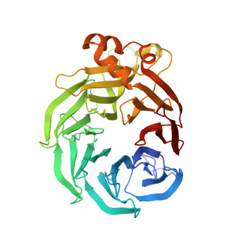
The inner workings of the hydrazine synthase multiprotein complex.

Dietl, A.Ferousi, C.Maalcke, W.J.Menzel, A.de Vries, S.Keltjens, J.T.Jetten, M.S.Kartal, B.Barends, T.R.

(2015) Nature 527: 394-397

  • DOI: https://doi.org/10.1038/nature15517
  • Primary Citation of Related Structures:  
    5C2V, 5C2W

  • PubMed Abstract: 

    Anaerobic ammonium oxidation (anammox) has a major role in the Earth's nitrogen cycle and is used in energy-efficient wastewater treatment. This bacterial process combines nitrite and ammonium to form dinitrogen (N2) gas, and has been estimated to synthesize up to 50% of the dinitrogen gas emitted into our atmosphere from the oceans. Strikingly, the anammox process relies on the highly unusual, extremely reactive intermediate hydrazine, a compound also used as a rocket fuel because of its high reducing power. So far, the enzymatic mechanism by which hydrazine is synthesized is unknown. Here we report the 2.7 Å resolution crystal structure, as well as biophysical and spectroscopic studies, of a hydrazine synthase multiprotein complex isolated from the anammox organism Kuenenia stuttgartiensis. The structure shows an elongated dimer of heterotrimers, each of which has two unique c-type haem-containing active sites, as well as an interaction point for a redox partner. Furthermore, a system of tunnels connects these active sites. The crystal structure implies a two-step mechanism for hydrazine synthesis: a three-electron reduction of nitric oxide to hydroxylamine at the active site of the γ-subunit and its subsequent condensation with ammonia, yielding hydrazine in the active centre of the α-subunit. Our results provide the first, to our knowledge, detailed structural insight into the mechanism of biological hydrazine synthesis, which is of major significance for our understanding of the conversion of nitrogenous compounds in nature.


  • Organizational Affiliation

    Department of Biomolecular Mechanisms, Max Planck Institute for Medical Research, 69120 Heidelberg, Germany.


Macromolecules
Find similar proteins by:  (by identity cutoff)  |  3D Structure
Entity ID: 1
MoleculeChains Sequence LengthOrganismDetailsImage
HYDRAZINE SYNTHASE ALPHA SUBUNIT
A, D
782Candidatus Kuenenia stuttgartiensisMutation(s): 0 
UniProt
Find proteins for Q1Q0T2 (Kuenenia stuttgartiensis)
Explore Q1Q0T2 
Go to UniProtKB:  Q1Q0T2
Entity Groups  
Sequence Clusters30% Identity50% Identity70% Identity90% Identity95% Identity100% Identity
UniProt GroupQ1Q0T2
Sequence Annotations
Expand
  • Reference Sequence
Find similar proteins by:  (by identity cutoff)  |  3D Structure
Entity ID: 2
MoleculeChains Sequence LengthOrganismDetailsImage
HYDRAZINE SYNTHASE BETA SUBUNIT
B, E
352Candidatus Kuenenia stuttgartiensisMutation(s): 0 
UniProt
Find proteins for Q1Q0T4 (Kuenenia stuttgartiensis)
Explore Q1Q0T4 
Go to UniProtKB:  Q1Q0T4
Entity Groups  
Sequence Clusters30% Identity50% Identity70% Identity90% Identity95% Identity100% Identity
UniProt GroupQ1Q0T4
Sequence Annotations
Expand
  • Reference Sequence
Find similar proteins by:  (by identity cutoff)  |  3D Structure
Entity ID: 3
MoleculeChains Sequence LengthOrganismDetailsImage
HYDRAZINE SYNTHASE GAMMA SUBUNIT
C, F
314Candidatus Kuenenia stuttgartiensisMutation(s): 0 
UniProt
Find proteins for Q1Q0T3 (Kuenenia stuttgartiensis)
Explore Q1Q0T3 
Go to UniProtKB:  Q1Q0T3
Entity Groups  
Sequence Clusters30% Identity50% Identity70% Identity90% Identity95% Identity100% Identity
UniProt GroupQ1Q0T3
Sequence Annotations
Expand
  • Reference Sequence
Small Molecules
Ligands 6 Unique
IDChains Name / Formula / InChI Key2D Diagram3D Interactions
HEC
Query on HEC

Download Ideal Coordinates CCD File 
CA [auth D]
DA [auth D]
K [auth A]
L [auth A]
LA [auth F]
CA [auth D],
DA [auth D],
K [auth A],
L [auth A],
LA [auth F],
MA [auth F],
V [auth C],
W [auth C]
HEME C
C34 H34 Fe N4 O4
HXQIYSLZKNYNMH-LJNAALQVSA-N
BET
Query on BET

Download Ideal Coordinates CCD File 
EA [auth D]
HA [auth E]
M [auth A]
N [auth A]
O [auth A]
EA [auth D],
HA [auth E],
M [auth A],
N [auth A],
O [auth A],
R [auth B],
X [auth C]
TRIMETHYL GLYCINE
C5 H12 N O2
KWIUHFFTVRNATP-UHFFFAOYSA-O
ZN
Query on ZN

Download Ideal Coordinates CCD File 
BA [auth D],
J [auth A]
ZINC ION
Zn
PTFCDOFLOPIGGS-UHFFFAOYSA-N
CA
Query on CA

Download Ideal Coordinates CCD File 
AA [auth D]
GA [auth E]
H [auth A]
I [auth A]
IA [auth F]
AA [auth D],
GA [auth E],
H [auth A],
I [auth A],
IA [auth F],
JA [auth F],
KA [auth F],
Q [auth B],
S [auth C],
T [auth C],
U [auth C],
Z [auth D]
CALCIUM ION
Ca
BHPQYMZQTOCNFJ-UHFFFAOYSA-N
CL
Query on CL

Download Ideal Coordinates CCD File 
G [auth A],
Y [auth D]
CHLORIDE ION
Cl
VEXZGXHMUGYJMC-UHFFFAOYSA-M
MG
Query on MG

Download Ideal Coordinates CCD File 
FA [auth E],
P [auth B]
MAGNESIUM ION
Mg
JLVVSXFLKOJNIY-UHFFFAOYSA-N
Experimental Data & Validation

Experimental Data

  • Method: X-RAY DIFFRACTION
  • Resolution: 2.70 Å
  • R-Value Free: 0.271 
  • R-Value Work: 0.234 
  • R-Value Observed: 0.236 
  • Space Group: H 3 2
Unit Cell:
Length ( Å )Angle ( ˚ )
a = 464.54α = 90
b = 464.54β = 90
c = 145.75γ = 120
Software Package:
Software NamePurpose
PHENIXrefinement
XDSdata reduction
XSCALEdata scaling
REFMACphasing

Structure Validation

View Full Validation Report


Ligand Structure Quality Assessment 


Entry History & Funding Information

Deposition Data


Funding OrganizationLocationGrant Number
NWONetherlands863.11.003
NWONetherlands142.16.1201
NWONetherlandsSpinozapremie
European Research CouncilNetherlandsERC232937

Revision History  (Full details and data files)

  • Version 1.0: 2015-10-14
    Type: Initial release
  • Version 1.1: 2015-11-04
    Changes: Database references
  • Version 1.2: 2015-12-02
    Changes: Database references
  • Version 2.0: 2023-11-15
    Changes: Atomic model, Author supporting evidence, Data collection, Database references, Derived calculations