5BYI | pdb_00005byi

Human carbonic anhydrase II with an azobenzene inhibitor (1d)


Experimental Data Snapshot

  • Method: X-RAY DIFFRACTION
  • Resolution: 1.15 Å
  • R-Value Free: 
    0.187 (Depositor), 0.181 (DCC) 
  • R-Value Work: 
    0.171 (Depositor), 0.165 (DCC) 
  • R-Value Observed: 
    0.174 (Depositor) 

wwPDB Validation 3D Report Full Report

Validation slider image for 5BYI

Ligand Structure Quality Assessment 


This is version 1.1 of the entry. See complete history

Literature

Azobenzene-based inhibitors of human carbonic anhydrase II.

Runtsch, L.S.Barber, D.M.Mayer, P.Groll, M.Trauner, D.Broichhagen, J.

(2015) Beilstein J Org Chem 11: 1129-1135

  • DOI: https://doi.org/10.3762/bjoc.11.127
  • Primary Citation Related Structures: 
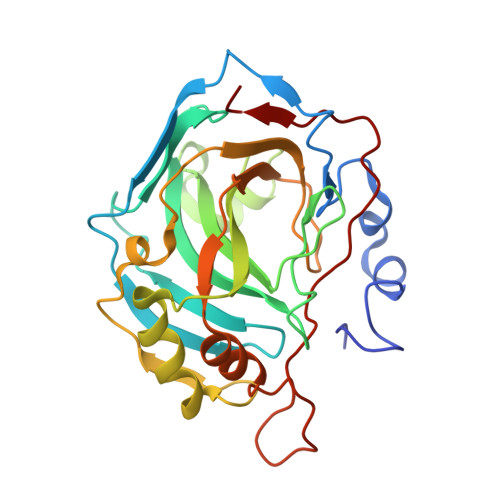
    5BYI

  • PubMed Abstract: 

    Aryl sulfonamides are a widely used drug class for the inhibition of carbonic anhydrases. In the context of our program of photochromic pharmacophores we were interested in the exploration of azobenzene-containing sulfonamides to block the catalytic activity of human carbonic anhydrase II (hCAII). Herein, we report the synthesis and in vitro evaluation of a small library of nine photochromic sulfonamides towards hCAII. All molecules are azobenzene-4-sulfonamides, which are substituted by different functional groups in the 4´-position and were characterized by X-ray crystallography. We aimed to investigate the influence of electron-donating or electron-withdrawing substituents on the inhibitory constant K i. With the aid of an hCAII crystal structure bound to one of the synthesized azobenzenes, we found that the electronic structure does not strongly affect inhibition. Taken together, all compounds are strong blockers of hCAII with K i = 25-65 nM that are potentially photochromic and thus combine studies from chemical synthesis, crystallography and enzyme kinetics.


  • Organizational Affiliation
    • Department of Chemistry, Ludwig-Maximilians-University Munich and Munich Center for Integrated Protein Science, Butenandtstrasse 5-13, 81377 Munich, Germany.

Macromolecule Content 

  • Total Structure Weight: 30.05 kDa 
  • Atom Count: 2,437 
  • Modeled Residue Count: 257 
  • Deposited Residue Count: 260 
  • Unique protein chains: 1

Macromolecules

Find similar proteins by:|  3D Structure
Entity ID: 1
MoleculeChains  Sequence LengthOrganismDetailsImage
Carbonic anhydrase 2260Homo sapiensMutation(s): 0 
Gene Names: CA2
EC: 4.2.1.1 (PDB Primary Data), 4.2.1.69 (UniProt)
UniProt & NIH Common Fund Data Resources
Find proteins for P00918 (Homo sapiens)
Explore P00918 
Go to UniProtKB:  P00918
PHAROS:  P00918
GTEx:  ENSG00000104267 
Entity Groups
Sequence Clusters30% Identity50% Identity70% Identity90% Identity95% Identity100% Identity
UniProt GroupP00918
Sequence Annotations
Expand
Reference Sequence

Small Molecules

Experimental Data & Validation

Experimental Data

  • Method: X-RAY DIFFRACTION
  • Resolution: 1.15 Å
  • R-Value Free:  0.187 (Depositor), 0.181 (DCC) 
  • R-Value Work:  0.171 (Depositor), 0.165 (DCC) 
  • R-Value Observed: 0.174 (Depositor) 
Space Group: P 1 21 1
Unit Cell:
Length ( Å )Angle ( ˚ )
a = 42.05α = 90
b = 41.22β = 104.23
c = 71.93γ = 90
Software Package:
Software NamePurpose
REFMACrefinement
XDSdata reduction
XDSdata scaling
REFMACphasing

Structure Validation

View Full Validation Report


Ligand Structure Quality Assessment 


Entry History 

Deposition Data

Revision History  (Full details and data files)

  • Version 1.0: 2015-08-05
    Type: Initial release
  • Version 1.1: 2024-05-08
    Changes: Data collection, Database references, Derived calculations