5BS4 | pdb_00005bs4

HIV-1 wild Type protease with GRL-047-11A (a methylamine bis-Tetrahydrofuran P2-Ligand, 4-amino sulfonamide derivative)


Experimental Data Snapshot

  • Method: X-RAY DIFFRACTION
  • Resolution: 1.29 Å
  • R-Value Free: 
    0.210 (Depositor), 0.192 (DCC) 
  • R-Value Work: 
    0.170 (Depositor), 0.155 (DCC) 
  • R-Value Observed: 
    0.172 (Depositor) 

Starting Model: experimental
View more details

wwPDB Validation 3D Report Full Report

Validation slider image for 5BS4

Ligand Structure Quality Assessment 


This is version 1.4 of the entry. See complete history

Literature

Design of HIV-1 Protease Inhibitors with Amino-bis-tetrahydrofuran Derivatives as P2-Ligands to Enhance Backbone-Binding Interactions: Synthesis, Biological Evaluation, and Protein-Ligand X-ray Studies.

Ghosh, A.K.Martyr, C.D.Osswald, H.L.Sheri, V.R.Kassekert, L.A.Chen, S.Agniswamy, J.Wang, Y.F.Hayashi, H.Aoki, M.Weber, I.T.Mitsuya, H.

(2015) J Med Chem 58: 6994-7006

  • DOI: https://doi.org/10.1021/acs.jmedchem.5b00900
  • Primary Citation Related Structures: 
    5BRY, 5BS4

  • PubMed Abstract: 

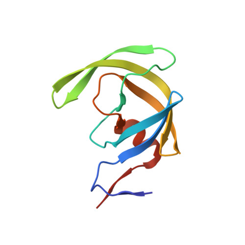
    Structure-based design, synthesis, and biological evaluation of a series of very potent HIV-1 protease inhibitors are described. In an effort to improve backbone ligand-binding site interactions, we have incorporated basic-amines at the C4 position of the bis-tetrahydrofuran (bis-THF) ring. We speculated that these substituents would make hydrogen bonding interactions in the flap region of HIV-1 protease. Synthesis of these inhibitors was performed diastereoselectively. A number of inhibitors displayed very potent enzyme inhibitory and antiviral activity. Inhibitors 25f, 25i, and 25j were evaluated against a number of highly-PI-resistant HIV-1 strains, and they exhibited improved antiviral activity over darunavir. Two high resolution X-ray structures of 25f- and 25g-bound HIV-1 protease revealed unique hydrogen bonding interactions with the backbone carbonyl group of Gly48 as well as with the backbone NH of Gly48 in the flap region of the enzyme active site. These ligand-binding site interactions are possibly responsible for their potent activity.


  • Organizational Affiliation
    • Department of Chemistry and Department of Medicinal Chemistry, Purdue University , West Lafayette, Indiana 47907, United States.

Macromolecule Content 

  • Total Structure Weight: 22.33 kDa 
  • Atom Count: 1,802 
  • Modeled Residue Count: 198 
  • Deposited Residue Count: 198 
  • Unique protein chains: 1

Macromolecules

Find similar proteins by:|  3D Structure
Entity ID: 1
MoleculeChains  Sequence LengthOrganismDetailsImage
Protease
A, B
99Human immunodeficiency virus type 1 BH10Mutation(s): 5 
Gene Names: gag-pol
EC: 3.4.23.16
UniProt
Find proteins for P03366 (Human immunodeficiency virus type 1 group M subtype B (isolate BH10))
Explore P03366 
Go to UniProtKB:  P03366
Entity Groups
Sequence Clusters30% Identity50% Identity70% Identity90% Identity95% Identity100% Identity
UniProt GroupP03366
Sequence Annotations
Expand
Reference Sequence

Small Molecules

Ligands 4 Unique
IDChains Name / Formula / InChI Key2D Diagram3D Interactions
4UX

Query on 4UX


Download:Ideal Coordinates CCD File
C [auth A](3R,3aS,4R,6aR)-4-(methylamino)hexahydrofuro[2,3-b]furan-3-yl [(2S,3R)-4-{[(4-aminophenyl)sulfonyl](2-methylpropyl)amino}-3-hydroxy-1-phenylbutan-2-yl]carbamate
C28 H40 N4 O7 S
ZQJLVCKJQHWULH-HNXVLALESA-N
ACT

Query on ACT


Download:Ideal Coordinates CCD File
F [auth A],
H [auth B],
I [auth B]
ACETATE ION
C2 H3 O2
QTBSBXVTEAMEQO-UHFFFAOYSA-M
CL

Query on CL


Download:Ideal Coordinates CCD File
E [auth A],
G [auth B]
CHLORIDE ION
Cl
VEXZGXHMUGYJMC-UHFFFAOYSA-M
NA

Query on NA


Download:Ideal Coordinates CCD File
D [auth A]SODIUM ION
Na
FKNQFGJONOIPTF-UHFFFAOYSA-N

Experimental Data & Validation

Experimental Data

  • Method: X-RAY DIFFRACTION
  • Resolution: 1.29 Å
  • R-Value Free:  0.210 (Depositor), 0.192 (DCC) 
  • R-Value Work:  0.170 (Depositor), 0.155 (DCC) 
  • R-Value Observed: 0.172 (Depositor) 
Space Group: P 21 21 2
Unit Cell:
Length ( Å )Angle ( ˚ )
a = 58.924α = 90
b = 85.987β = 90
c = 46.138γ = 90
Software Package:
Software NamePurpose
HKL-2000data reduction
HKL-2000data scaling
PHASERphasing
SHELXL-97refinement

Structure Validation

View Full Validation Report


Ligand Structure Quality Assessment 


Entry History 

& Funding Information

Deposition Data


Funding OrganizationLocationGrant Number
National Institutes of Health/National Institute of General Medical Sciences (NIH/NIGMS)United StatesGM53386
National Institutes of Health/National Institute of General Medical Sciences (NIH/NIGMS)United StatesGM62920
Department of Energy (DOE, United States)United StatesW-31-109-Eng-38
National Institutes of Health/National Cancer Institute (NIH/NCI)United Statesby the Intramural Research Program of the Center for Cancer Research
the Ministry of Education, Culture, Sports, Science, and TechnologyJapanby a Grant-in-Aid for Scientific Research (Priority Areas)
the Ministry of Education, Culture, Sports, Science, and TechnologyJapanthe Grant to the Cooperative Research Project on Clinical and Epidemiological Studies of Emerging and Reemerging Infectious Diseases (Renkei Jigyo)
Ministry of Health, Welfare, and LaborJapanGrant for Promotion of AIDS Research

Revision History  (Full details and data files)

  • Version 1.0: 2015-09-09
    Type: Initial release
  • Version 1.1: 2015-09-23
    Changes: Database references
  • Version 1.2: 2017-09-20
    Changes: Author supporting evidence, Database references, Derived calculations
  • Version 1.3: 2019-12-04
    Changes: Author supporting evidence
  • Version 1.4: 2023-09-27
    Changes: Data collection, Database references, Derived calculations, Refinement description