Mutations in adenine-binding pockets enhance catalytic properties of NAD(P)H-dependent enzymes.
Cahn, J.K., Baumschlager, A., Brinkmann-Chen, S., Arnold, F.H.(2016) Protein Eng Des Sel 29: 31-38
- PubMed: 26512129 Search on PubMedSearch on PubMed Central
- DOI: https://doi.org/10.1093/protein/gzv057
- Primary Citation Related Structures:
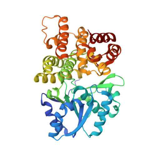
5BR4 - PubMed Abstract:
NAD(P)H-dependent enzymes are ubiquitous in metabolism and cellular processes and are also of great interest for pharmaceutical and industrial applications. Here, we present a structure-guided enzyme engineering strategy for improving catalytic properties of NAD(P)H-dependent enzymes toward native or native-like reactions using mutations to the enzyme's adenine-binding pocket, distal to the site of catalysis. Screening single-site saturation mutagenesis libraries identified mutations that increased catalytic efficiency up to 10-fold in 7 out of 10 enzymes. The enzymes improved in this study represent three different cofactor-binding folds (Rossmann, DHQS-like, and FAD/NAD binding) and utilize both NADH and NADPH. Structural and biochemical analyses show that the improved activities are accompanied by minimal changes in other properties (cooperativity, thermostability, pH optimum, uncoupling), and initial tests on two enzymes (ScADH6 and EcFucO) show improved functionality in Escherichia coli.
- Division of Chemistry and Chemical Engineering, California Institute of Technology, 1200 E California Blvd, MC 210-41, Pasadena, CA 91125, USA.
Organizational Affiliation: