5AWH | pdb_00005awh

Rhodobacter sphaeroides Argonaute in complex with guide RNA/target DNA heteroduplex


Experimental Data Snapshot

  • Method: X-RAY DIFFRACTION
  • Resolution: 2.00 Å
  • R-Value Free: 
    0.233 (Depositor), 0.241 (DCC) 
  • R-Value Work: 
    0.184 (Depositor), 0.194 (DCC) 
  • R-Value Observed: 
    0.187 (Depositor) 

wwPDB Validation 3D Report Full Report

Validation slider image for 5AWH

This is version 1.2 of the entry. See complete history

Literature

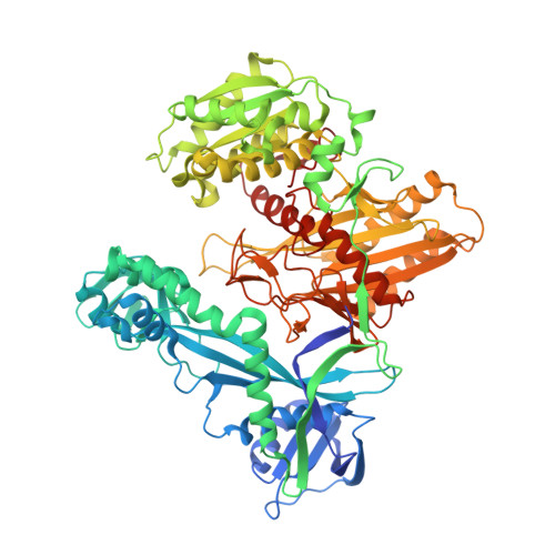
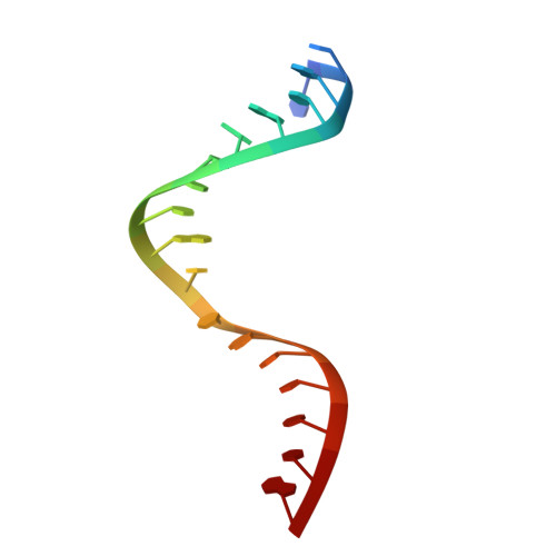
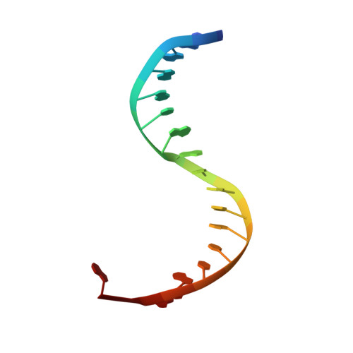
Structural basis for the recognition of guide RNA and target DNA heteroduplex by Argonaute.

Miyoshi, T.Ito, K.Murakami, R.Uchiumi, T.

(2016) Nat Commun 7: 11846-11846

  • DOI: https://doi.org/10.1038/ncomms11846
  • Primary Citation Related Structures: 
    5AWH

  • PubMed Abstract: 

    Argonaute proteins are key players in the gene silencing mechanisms mediated by small nucleic acids in all domains of life from bacteria to eukaryotes. However, little is known about the Argonaute protein that recognizes guide RNA/target DNA. Here, we determine the 2 Å crystal structure of Rhodobacter sphaeroides Argonaute (RsAgo) in a complex with 18-nucleotide guide RNA and its complementary target DNA. The heteroduplex maintains Watson-Crick base-pairing even in the 3'-region of the guide RNA between the N-terminal and PIWI domains, suggesting a recognition mode by RsAgo for stable interaction with the target strand. In addition, the MID/PIWI interface of RsAgo has a system that specifically recognizes the 5' base-U of the guide RNA, and the duplex-recognition loop of the PAZ domain is important for the DNA silencing activity. Furthermore, we show that Argonaute discriminates the nucleic acid type (RNA/DNA) by recognition of the duplex structure of the seed region.


  • Organizational Affiliation
    • Center for Transdisciplinary Research, Niigata University, 8050 Ikarashi 2-no-cho, Nishi-ku, Niigata 950-2181, Japan.

Macromolecule Content 

  • Total Structure Weight: 196.83 kDa 
  • Atom Count: 13,805 
  • Modeled Residue Count: 1,546 
  • Deposited Residue Count: 1,628 
  • Unique protein chains: 1
  • Unique nucleic acid chains: 2

Macromolecules


Find similar proteins by:|  3D Structure
Entity ID: 1
MoleculeChains  Sequence LengthOrganismDetailsImage
Uncharacterized protein
A, B
778Cereibacter sphaeroides ATCC 17025Mutation(s): 0 
Gene Names: Rsph17025_3694
UniProt
Find proteins for A4WYU7 (Cereibacter sphaeroides (strain ATCC 17025 / ATH 2.4.3))
Explore A4WYU7 
Go to UniProtKB:  A4WYU7
Entity Groups
Sequence Clusters30% Identity50% Identity70% Identity90% Identity95% Identity100% Identity
UniProt GroupA4WYU7
Sequence Annotations
Expand
Reference Sequence
Find similar nucleic acids by:  Sequence
Entity ID: 2
MoleculeChains LengthOrganismImage
RNA (5'-D(P*UP*UP*AP*CP*AP*AP*CP*CP*UP*AP*CP*UP*AP*CP*CP*UP*CP*G)-3')
C, E
18synthetic construct
Sequence Annotations
Expand
Reference Sequence
Find similar nucleic acids by:  Sequence
Entity ID: 3
MoleculeChains LengthOrganismImage
DNA (5'-D(*CP*GP*AP*GP*GP*TP*AP*GP*TP*AP*GP*GP*TP*TP*GP*TP*AP*A)-3')
D, F
18synthetic construct
Sequence Annotations
Expand
Reference Sequence

Experimental Data & Validation

Experimental Data

  • Method: X-RAY DIFFRACTION
  • Resolution: 2.00 Å
  • R-Value Free:  0.233 (Depositor), 0.241 (DCC) 
  • R-Value Work:  0.184 (Depositor), 0.194 (DCC) 
  • R-Value Observed: 0.187 (Depositor) 
Space Group: P 1 21 1
Unit Cell:
Length ( Å )Angle ( ˚ )
a = 68.186α = 90
b = 118.303β = 95.83
c = 118.48γ = 90
Software Package:
Software NamePurpose
HKL-2000data scaling
REFMACrefinement
PDB_EXTRACTdata extraction
SHARPphasing
HKL-2000data reduction

Structure Validation

View Full Validation Report


Entry History 

Deposition Data

Revision History  (Full details and data files)

  • Version 1.0: 2016-07-06
    Type: Initial release
  • Version 1.1: 2020-02-26
    Changes: Data collection, Derived calculations
  • Version 1.2: 2024-03-20
    Changes: Data collection, Database references, Derived calculations