5AQD

Crystal structure of Phormidium Phycoerythrin at pH 8.5


Experimental Data Snapshot

  • Method: X-RAY DIFFRACTION
  • Resolution: 2.12 Å
  • R-Value Free: 0.239 
  • R-Value Work: 0.175 
  • R-Value Observed: 0.178 

wwPDB Validation   3D Report Full Report


Ligand Structure Quality Assessment 


This is version 1.2 of the entry. See complete history


Literature

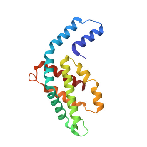
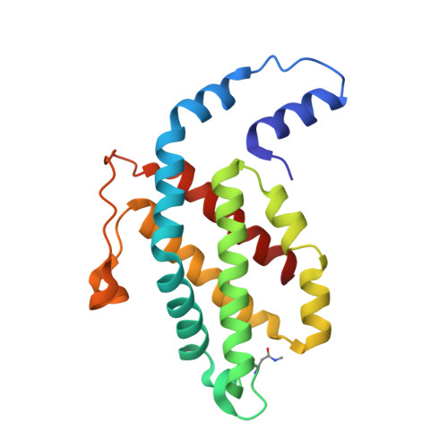
Crystal Structure Analysis of C-Phycoerythrin from Marine Cyanobacterium Phormidium Sp. A09Dm.

Kumar, V.Sonani, R.R.Sharma, M.Gupta, G.D.Madamwar, D.

(2016) Photosynth Res 129: 17

  • DOI: https://doi.org/10.1007/s11120-016-0259-5
  • Primary Citation of Related Structures:  
    5AQD, 5FVB

  • PubMed Abstract: 

    The role of unique sequence features of C-phycoerythrin, isolated from Phormidium sp. A09DM, has been investigated by crystallographic studies. Two conserved indels (i.e. inserts or deletions) are found in the β-subunit of Phormidium phycoerythrin that are distinctive characteristics of large number of cyanobacterial sequences. The identified signatures are a two-residue deletion from position 21 and a nine-residue insertion at position 146. Crystals of Phormidium phycoerythrin were obtained at pH values of 5 and 8.5, and structures have been resolved to high precision at 1.95 and 2.1 Å resolution, respectively. In both the structures, heterodimers of α- and β- subunits assemble as hexamers. The 7-residue insertion at position 146 significantly reduces solvent exposure of π-conjugated A-C rings of a phycoerythrobilin (PEB) chromophore, and can influence energy absorption and energy transfer characteristics. The structural analyses (with 12-fold redundancy) suggest that protein micro-environment alone dictates the conformation of bound chromophores. The low- and high-energy absorbing chromophores are identified based on A-B ring coplanarity. The spatial distribution of these is found to be similar to that observed in R-phycoerythrin, suggesting the direction of energy transfer from outer-surface of hexamer to inner-hollow cavity in the Phormidium protein. The crystal structures also reveal that a commonly observed Hydrogen-bonding network in phycobiliproteins, involving chromophore bound to α-subunit and amino acid at position 73 of β-subunit, may not be essential for structural and functional integrity of C-phycoerythrin orthologs. In solution, the protein displays slight red shift and decrease in fluorescence emission at acidic pH. The mechanism for which may be static and correlates with the proximity of +ve electric field of Arg148 to the C-ring of a PEB chromophore.


  • Organizational Affiliation

    Protein Crystallography Section, Solid State Physics Division, Bhabha Atomic Research Centre, Mumbai, 400085, India. vinay@barc.gov.in.


Macromolecules
Find similar proteins by:  (by identity cutoff)  |  3D Structure
Entity ID: 1
MoleculeChains Sequence LengthOrganismDetailsImage
PHYCOERYTHRIN ALPHA SUBUNIT
A, B, C, D, E
A, B, C, D, E, F, G, H, I, J, K, L
164Phormidium rubidum A09DMMutation(s): 0 
UniProt
Find proteins for A0A0E3W010 (Phormidium rubidum A09DM)
Explore A0A0E3W010 
Go to UniProtKB:  A0A0E3W010
Entity Groups  
Sequence Clusters30% Identity50% Identity70% Identity90% Identity95% Identity100% Identity
UniProt GroupA0A0E3W010
Sequence Annotations
Expand
  • Reference Sequence
Find similar proteins by:  (by identity cutoff)  |  3D Structure
Entity ID: 2
MoleculeChains Sequence LengthOrganismDetailsImage
PHYCOERYTHRIN BETA SUBUNIT
M, N, O, P, Q
M, N, O, P, Q, R, S, T, U, V, W, X
184Phormidium rubidum A09DMMutation(s): 0 
UniProt
Find proteins for A0A0E4G455 (Phormidium rubidum A09DM)
Explore A0A0E4G455 
Go to UniProtKB:  A0A0E4G455
Entity Groups  
Sequence Clusters30% Identity50% Identity70% Identity90% Identity95% Identity100% Identity
UniProt GroupA0A0E4G455
Sequence Annotations
Expand
  • Reference Sequence
Small Molecules
Ligands 3 Unique
IDChains Name / Formula / InChI Key2D Diagram3D Interactions
PEB
Query on PEB

Download Ideal Coordinates CCD File 
AB [auth I]
AC [auth P]
AD [auth V]
BB [auth I]
BC [auth P]
AB [auth I],
AC [auth P],
AD [auth V],
BB [auth I],
BC [auth P],
CA [auth B],
CC [auth P],
CD [auth W],
DA [auth B],
DD [auth W],
EB [auth J],
EC [auth Q],
ED [auth W],
FA [auth C],
FB [auth J],
FC [auth Q],
GA [auth C],
GC [auth Q],
GD [auth X],
HB [auth K],
HD [auth X],
IA [auth D],
IB [auth K],
IC [auth R],
ID [auth X],
JA [auth D],
JC [auth R],
KB [auth L],
KC [auth R],
LB [auth L],
MA [auth E],
MC [auth S],
NA [auth E],
NC [auth S],
OB [auth M],
OC [auth S],
PB [auth M],
QA [auth F],
QB [auth M],
QC [auth T],
RA [auth F],
RC [auth T],
SB [auth N],
SC [auth T],
TA [auth G],
TB [auth N],
UA [auth G],
UB [auth N],
UC [auth U],
VC [auth U],
WA [auth H],
WB [auth O],
WC [auth U],
XA [auth H],
XB [auth O],
Y [auth A],
YB [auth O],
YC [auth V],
Z [auth A],
ZC [auth V]
PHYCOERYTHROBILIN
C33 H40 N4 O6
NKCBCVIFPXGHAV-WAVSMFBNSA-N
SO4
Query on SO4

Download Ideal Coordinates CCD File 
BA [auth A]
BD [auth V]
DB [auth I]
DC [auth P]
FD [auth W]
BA [auth A],
BD [auth V],
DB [auth I],
DC [auth P],
FD [auth W],
HC [auth Q],
JD [auth X],
LA [auth D],
LC [auth R],
NB [auth L],
PA [auth E],
PC [auth S],
RB [auth M],
TC [auth T],
VB [auth N],
XC [auth U],
ZA [auth H],
ZB [auth O]
SULFATE ION
O4 S
QAOWNCQODCNURD-UHFFFAOYSA-L
GOL
Query on GOL

Download Ideal Coordinates CCD File 
AA [auth A]
CB [auth I]
EA [auth B]
GB [auth J]
HA [auth C]
AA [auth A],
CB [auth I],
EA [auth B],
GB [auth J],
HA [auth C],
JB [auth K],
KA [auth D],
MB [auth L],
OA [auth E],
SA [auth F],
VA [auth G],
YA [auth H]
GLYCEROL
C3 H8 O3
PEDCQBHIVMGVHV-UHFFFAOYSA-N
Modified Residues  1 Unique
IDChains TypeFormula2D DiagramParent
MEN
Query on MEN
M, N, O, P, Q
M, N, O, P, Q, R, S, T, U, V, W, X
L-PEPTIDE LINKINGC5 H10 N2 O3ASN
Experimental Data & Validation

Experimental Data

  • Method: X-RAY DIFFRACTION
  • Resolution: 2.12 Å
  • R-Value Free: 0.239 
  • R-Value Work: 0.175 
  • R-Value Observed: 0.178 
  • Space Group: P 1
Unit Cell:
Length ( Å )Angle ( ˚ )
a = 108.298α = 78.94
b = 108.36β = 82.5
c = 116.592γ = 60.34
Software Package:
Software NamePurpose
PHENIXrefinement
autoPROCdata reduction
XDSdata reduction
Aimlessdata scaling
PHASERphasing

Structure Validation

View Full Validation Report


Ligand Structure Quality Assessment 


Entry History 

Deposition Data

Revision History  (Full details and data files)

  • Version 1.0: 2016-06-29
    Type: Initial release
  • Version 1.1: 2018-01-31
    Changes: Data collection
  • Version 1.2: 2024-01-10
    Changes: Data collection, Database references, Derived calculations, Other, Refinement description