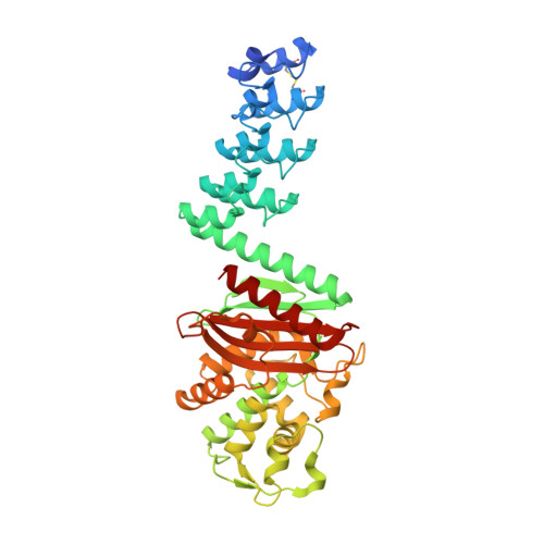
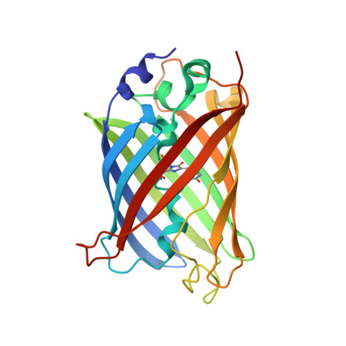
5AQB

DARPin-based Crystallization Chaperones exploit Molecular Geometry as a Screening Dimension in Protein Crystallography


Experimental Data Snapshot

  • Method: X-RAY DIFFRACTION
  • Resolution: 1.37 Å
  • R-Value Free: 0.177 
  • R-Value Work: 0.156 
  • R-Value Observed: 0.157 

Starting Models: experimental
View more details

wwPDB Validation   3D Report Full Report


This is version 3.1 of the entry. See complete history


Literature

Darpin-Based Crystallization Chaperones Exploit Molecular Geometry as a Screening Dimension in Protein Crystallography

Batyuk, A.Wu, Y.Honegger, A.Heberling, M.Plueckthun, A.

(2016) J Mol Biol 428: 1574

  • DOI: https://doi.org/10.1016/j.jmb.2016.03.002
  • Primary Citation of Related Structures:  
    5AQ7, 5AQ8, 5AQ9, 5AQA, 5AQB

  • PubMed Abstract: 

    DARPin libraries, based on a Designed Ankyrin Repeat Protein consensus framework, are a rich source of binding partners for a wide variety of proteins. Their modular structure, stability, ease of in vitro selection and high production yields make DARPins an ideal starting point for further engineering. The X-ray structures of around 30 different DARPin complexes demonstrate their ability to facilitate crystallization of their target proteins by restricting flexibility and preventing undesired interactions of the target molecule. However, their small size (18 kDa), very hydrophilic surface and repetitive structure can limit the DARPins' ability to provide essential crystal contacts and their usefulness as a search model for addressing the crystallographic phase problem in molecular replacement. To optimize DARPins for their application as crystallization chaperones, rigid domain-domain fusions of the DARPins to larger proteins, proven to yield high-resolution crystal structures, were generated. These fusions were designed in such a way that they affect only one of the terminal capping repeats of the DARPin and do not interfere with residues involved in target binding, allowing to exchange at will the binding specificities of the DARPin in the fusion construct. As a proof of principle, we designed rigid fusions of a stabilized version of Escherichia coli TEM-1 β-lactamase to the C-terminal capping repeat of various DARPins in six different relative domain orientations. Five crystal structures representing four different fusion constructs, alone or in complex with the cognate target, show the predicted relative domain orientations and prove the validity of the concept.


  • Organizational Affiliation

    Department of Biochemistry, University of Zurich, Winterthurerstrasse 190, CH-8057 Zürich, Switzerland.


Macromolecules
Find similar proteins by:  (by identity cutoff)  |  3D Structure
Entity ID: 1
MoleculeChains Sequence LengthOrganismDetailsImage
3G61_DB15V4428synthetic constructMutation(s): 0 
Entity Groups  
Sequence Clusters30% Identity50% Identity70% Identity90% Identity95% Identity100% Identity
Sequence Annotations
Expand
  • Reference Sequence
Find similar proteins by:  (by identity cutoff)  |  3D Structure
Entity ID: 2
MoleculeChains Sequence LengthOrganismDetailsImage
GREEN FLUORESCENT PROTEIN229Aequorea victoriaMutation(s): 1 
UniProt
Find proteins for P42212 (Aequorea victoria)
Explore P42212 
Go to UniProtKB:  P42212
Entity Groups  
Sequence Clusters30% Identity50% Identity70% Identity90% Identity95% Identity100% Identity
UniProt GroupP42212
Sequence Annotations
Expand
  • Reference Sequence
Small Molecules
Modified Residues  1 Unique
IDChains TypeFormula2D DiagramParent
CRO
Query on CRO
B
L-PEPTIDE LINKINGC15 H17 N3 O5THR, TYR, GLY
Experimental Data & Validation

Experimental Data

  • Method: X-RAY DIFFRACTION
  • Resolution: 1.37 Å
  • R-Value Free: 0.177 
  • R-Value Work: 0.156 
  • R-Value Observed: 0.157 
  • Space Group: C 1 2 1
Unit Cell:
Length ( Å )Angle ( ˚ )
a = 90.15α = 90
b = 96.17β = 118.99
c = 92.78γ = 90
Software Package:
Software NamePurpose
PHENIXrefinement
XDSdata reduction
XSCALEdata scaling
PHASERphasing

Structure Validation

View Full Validation Report


Entry History 

Deposition Data

Revision History  (Full details and data files)

  • Version 1.0: 2016-03-23
    Type: Initial release
  • Version 1.1: 2016-04-06
    Changes: Database references
  • Version 1.2: 2016-05-18
    Changes: Database references
  • Version 2.0: 2019-10-23
    Changes: Atomic model, Data collection, Database references, Derived calculations, Other
  • Version 3.0: 2023-11-15
    Changes: Advisory, Atomic model, Data collection, Database references
  • Version 3.1: 2024-01-10
    Changes: Refinement description