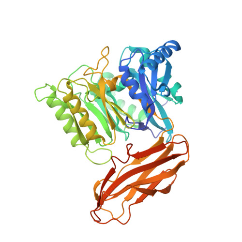
5AK8

Structure of C351A mutant of Porphyromonas gingivalis peptidylarginine deiminase


Experimental Data Snapshot

  • Method: X-RAY DIFFRACTION
  • Resolution: 1.48 Å
  • R-Value Free: 0.164 
  • R-Value Work: 0.127 
  • R-Value Observed: 0.129 

wwPDB Validation   3D Report Full Report


Ligand Structure Quality Assessment 


This is version 1.5 of the entry. See complete history


Literature

Crystal Structure of Porphyromonas Gingivalis Peptidylarginine Deiminase: Implications for Autoimmunity in Rheumatoid Arthritis.

Montgomery, A.B.Kopec, J.Shrestha, L.Thezenas, M.L.Burgess-Brown, N.A.Fischer, R.Yue, W.W.Venables, P.J.

(2016) Ann Rheum Dis 75: 1255

  • DOI: https://doi.org/10.1136/annrheumdis-2015-207656
  • Primary Citation of Related Structures:  
    5AK7, 5AK8

  • PubMed Abstract: 

    Periodontitis (PD) is a known risk factor for rheumatoid arthritis (RA) and there is increasing evidence that the link between the two diseases is due to citrullination by the unique bacterial peptidylarginine deiminase (PAD) enzyme expressed by periodontal pathogen Pophyromonas gingivalis (PPAD). However, the precise mechanism by which PPAD could generate potentially immunogenic peptides has remained controversial due to lack of information about the structural and catalytic mechanisms of the enzyme. By solving the 3D structure of PPAD we aim to characterise activity and elucidate potential mechanisms involved in breach of tolerance to citrullinated proteins in RA. PPAD and a catalytically inactive mutant PPAD(C351A) were crystallised and their 3D structures solved. Key residues identified from 3D structures were examined by mutations. Fibrinogen and α-enolase were incubated with PPAD and P. gingivalis arginine gingipain (RgpB) and citrullinated peptides formed were sequenced and quantified by mass spectrometry. Here, we solve the crystal structure of a truncated, highly active form of PPAD. We confirm catalysis is mediated by the following residues: Asp130, His236, Asp238, Asn297 and Cys351 and show Arg152 and Arg154 may determine the substrate specificity of PPAD for C-terminal arginines. We demonstrate the formation of 37 C-terminally citrullinated peptides from fibrinogen and 11 from α-enolase following incubation with tPPAD and RgpB. PPAD displays an unequivocal specificity for C-terminal arginine residues and readily citrullinates peptides from key RA autoantigens. The formation of these novel citrullinated peptides may be involved in breach of tolerance to citrullinated proteins in RA.


  • Organizational Affiliation

    Kennedy institute of Rheumatology, Nuffield Department of Orthopaedics, Rheumatology and Musculoskeletal Sciences, University of Oxford, Oxford, UK.


Macromolecules
Find similar proteins by:  (by identity cutoff)  |  3D Structure
Entity ID: 1
MoleculeChains Sequence LengthOrganismDetailsImage
PEPTIDYLARGININE DEIMINASE438Porphyromonas gingivalisMutation(s): 1 
EC: 3.5.3
UniProt
Find proteins for Q9RQJ2 (Porphyromonas gingivalis (strain ATCC BAA-308 / W83))
Explore Q9RQJ2 
Go to UniProtKB:  Q9RQJ2
Entity Groups  
Sequence Clusters30% Identity50% Identity70% Identity90% Identity95% Identity100% Identity
UniProt GroupQ9RQJ2
Sequence Annotations
Expand
  • Reference Sequence
Small Molecules
Ligands 4 Unique
IDChains Name / Formula / InChI Key2D Diagram3D Interactions
ARG
Query on ARG

Download Ideal Coordinates CCD File 
C [auth A]ARGININE
C6 H15 N4 O2
ODKSFYDXXFIFQN-BYPYZUCNSA-O
ALA
Query on ALA

Download Ideal Coordinates CCD File 
B [auth A]ALANINE
C3 H7 N O2
QNAYBMKLOCPYGJ-REOHCLBHSA-N
EDO
Query on EDO

Download Ideal Coordinates CCD File 
E [auth A]
F [auth A]
G [auth A]
H [auth A]
I [auth A]
E [auth A],
F [auth A],
G [auth A],
H [auth A],
I [auth A],
J [auth A],
K [auth A],
L [auth A],
M [auth A],
N [auth A],
O [auth A],
P [auth A],
Q [auth A],
R [auth A],
S [auth A],
T [auth A],
U [auth A],
V [auth A]
1,2-ETHANEDIOL
C2 H6 O2
LYCAIKOWRPUZTN-UHFFFAOYSA-N
NA
Query on NA

Download Ideal Coordinates CCD File 
D [auth A]SODIUM ION
Na
FKNQFGJONOIPTF-UHFFFAOYSA-N
Experimental Data & Validation

Experimental Data

  • Method: X-RAY DIFFRACTION
  • Resolution: 1.48 Å
  • R-Value Free: 0.164 
  • R-Value Work: 0.127 
  • R-Value Observed: 0.129 
  • Space Group: C 1 2 1
Unit Cell:
Length ( Å )Angle ( ˚ )
a = 101.79α = 90
b = 84.09β = 92.64
c = 55.46γ = 90
Software Package:
Software NamePurpose
REFMACrefinement
PHASERphasing

Structure Validation

View Full Validation Report


Ligand Structure Quality Assessment 


Entry History 

Deposition Data

Revision History  (Full details and data files)

  • Version 1.0: 2015-07-22
    Type: Initial release
  • Version 1.1: 2015-08-05
    Changes: Database references
  • Version 1.2: 2015-08-19
    Changes: Structure summary
  • Version 1.3: 2016-05-25
    Changes: Database references
  • Version 1.4: 2018-06-20
    Changes: Advisory, Data collection, Derived calculations
  • Version 1.5: 2024-05-08
    Changes: Data collection, Database references, Derived calculations, Other