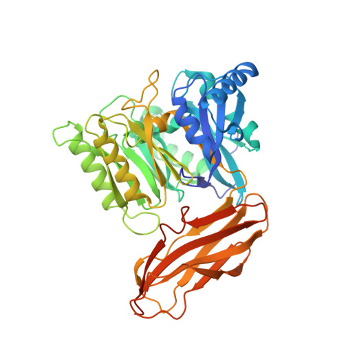
Crystal Structure of Porphyromonas Gingivalis Peptidylarginine Deiminase: Implications for Autoimmunity in Rheumatoid Arthritis.
Montgomery, A.B., Kopec, J., Shrestha, L., Thezenas, M.L., Burgess-Brown, N.A., Fischer, R., Yue, W.W., Venables, P.J.(2016) Ann Rheum Dis 75: 1255
- PubMed: 26209657 Search on PubMedSearch on PubMed Central
- DOI: https://doi.org/10.1136/annrheumdis-2015-207656
- Primary Citation Related Structures:
5AK7, 5AK8 - PubMed Abstract:
Periodontitis (PD) is a known risk factor for rheumatoid arthritis (RA) and there is increasing evidence that the link between the two diseases is due to citrullination by the unique bacterial peptidylarginine deiminase (PAD) enzyme expressed by periodontal pathogen Pophyromonas gingivalis (PPAD). However, the precise mechanism by which PPAD could generate potentially immunogenic peptides has remained controversial due to lack of information about the structural and catalytic mechanisms of the enzyme. By solving the 3D structure of PPAD we aim to characterise activity and elucidate potential mechanisms involved in breach of tolerance to citrullinated proteins in RA. PPAD and a catalytically inactive mutant PPAD(C351A) were crystallised and their 3D structures solved. Key residues identified from 3D structures were examined by mutations. Fibrinogen and α-enolase were incubated with PPAD and P. gingivalis arginine gingipain (RgpB) and citrullinated peptides formed were sequenced and quantified by mass spectrometry. Here, we solve the crystal structure of a truncated, highly active form of PPAD. We confirm catalysis is mediated by the following residues: Asp130, His236, Asp238, Asn297 and Cys351 and show Arg152 and Arg154 may determine the substrate specificity of PPAD for C-terminal arginines. We demonstrate the formation of 37 C-terminally citrullinated peptides from fibrinogen and 11 from α-enolase following incubation with tPPAD and RgpB. PPAD displays an unequivocal specificity for C-terminal arginine residues and readily citrullinates peptides from key RA autoantigens. The formation of these novel citrullinated peptides may be involved in breach of tolerance to citrullinated proteins in RA.
- Kennedy institute of Rheumatology, Nuffield Department of Orthopaedics, Rheumatology and Musculoskeletal Sciences, University of Oxford, Oxford, UK.
Organizational Affiliation: