


Dyrk1A with Ldn-211898
Elkins, J.M., Soundararajan, M., Muniz, J.R.C., Cuny, G., Higgins, J., Edwards, A., Bountra, C., Knapp, S.To be published.
Experimental Data Snapshot
Starting Model: experimental
View more details
Entity ID: 1 | |||||
|---|---|---|---|---|---|
| Molecule | Chains | Sequence Length | Organism | Details | Image |
| DYRK1A DUAL-SPECIFICITY TYROSINE-PHOSPHORYLATION REGULATED KINASE 1A | 382 | Homo sapiens | Mutation(s): 0 EC: 2.7.11.1 (PDB Primary Data), 2.7.12.1 (UniProt), 2.7.11.23 (UniProt) | 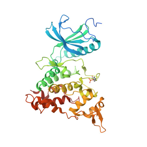 | |
UniProt & NIH Common Fund Data Resources | |||||
PHAROS: Q13627 GTEx: ENSG00000157540 | |||||
Entity Groups | |||||
| Sequence Clusters | 30% Identity50% Identity70% Identity90% Identity95% Identity100% Identity | ||||
| UniProt Group | Q13627 | ||||
Sequence AnnotationsExpand | |||||
Reference Sequence | |||||
| Ligands 2 Unique | |||||
|---|---|---|---|---|---|
| ID | Chains | Name / Formula / InChI Key | 2D Diagram | 3D Interactions | |
| AWR Download:Ideal Coordinates CCD File | E [auth A], H [auth B], L [auth C], O [auth D] | 4-(7-METHOXY-1-(TRIFLUOROMETHYL)-9H-PYRIDO[3,4-B]INDOL-9-yl)butan-1-amine C17 H18 F3 N3 O JVBWXORXTBDUMH-UHFFFAOYSA-N |  | ||
| PO4 Download:Ideal Coordinates CCD File | F [auth A] G [auth A] I [auth B] J [auth B] K [auth B] | PHOSPHATE ION O4 P NBIIXXVUZAFLBC-UHFFFAOYSA-K |  | ||
| Modified Residues 1 Unique | |||||
|---|---|---|---|---|---|
| ID | Chains | Type | Formula | 2D Diagram | Parent |
| PTR Query on PTR | A, B, C, D | L-PEPTIDE LINKING | C9 H12 N O6 P |  | TYR |
| Length ( Å ) | Angle ( ˚ ) |
|---|---|
| a = 89.955 | α = 90 |
| b = 90.891 | β = 90 |
| c = 232.468 | γ = 90 |
| Software Name | Purpose |
|---|---|
| REFMAC | refinement |
| MOSFLM | data reduction |
| SCALA | data scaling |
| PHASER | phasing |


