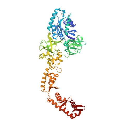
4ZU9 | pdb_00004zu9

Crystal structure of bacterial selenocysteine-specific elongation factor EF-Sec


Experimental Data Snapshot

  • Method: X-RAY DIFFRACTION
  • Resolution: 3.19 Å
  • R-Value Free: 
    0.261 (Depositor), 0.264 (DCC) 
  • R-Value Work: 
    0.206 (Depositor), 0.213 (DCC) 
  • R-Value Observed: 
    0.209 (Depositor) 

wwPDB Validation   3D Report Full Report


Ligand Structure Quality Assessment 

Created with Raphaël 2.3.0Worse 01 BetterLigand structure goodness of fit to experimental dataBest fitted GNPClick on this verticalbar to view details

This is version 1.3 of the entry. See complete history


Literature

Crystal structure of the full-length bacterial selenocysteine-specific elongation factor SelB

Itoh, Y.Sekine, S.Yokoyama, S.

(2015) Nucleic Acids Res 43: 9028-9038

  • DOI: https://doi.org/10.1093/nar/gkv833
  • Primary Citation of Related Structures:  
    4ZU9

  • PubMed Abstract: 

    Selenocysteine (Sec), the 21(st) amino acid in translation, uses its specific tRNA (tRNA(Sec)) to recognize the UGA codon. The Sec-specific elongation factor SelB brings the selenocysteinyl-tRNA(Sec) (Sec-tRNA(Sec)) to the ribosome, dependent on both an in-frame UGA and a Sec-insertion sequence (SECIS) in the mRNA. The bacterial SelB binds mRNA through its C-terminal region, for which crystal structures have been reported. In this study, we determined the crystal structure of the full-length SelB from the bacterium Aquifex aeolicus, in complex with a GTP analog, at 3.2-Å resolution. SelB consists of three EF-Tu-like domains (D1-3), followed by four winged-helix domains (WHD1-4). The spacer region, connecting the N- and C-terminal halves, fixes the position of WHD1 relative to D3. The binding site for the Sec moiety of Sec-tRNA(Sec) is located on the interface between D1 and D2, where a cysteine molecule from the crystallization solution is coordinated by Arg residues, which may mimic Sec binding. The Sec-binding site is smaller and more exposed than the corresponding site of EF-Tu. Complex models of Sec-tRNA(Sec), SECIS RNA, and the 70S ribosome suggest that the unique secondary structure of tRNA(Sec) allows SelB to specifically recognize tRNA(Sec) and characteristically place it at the ribosomal A-site.


  • Organizational Affiliation

    RIKEN Systems and Structural Biology Center, 1-7-22 Suehiro-cho, Tsurumi, Yokohama 230-0045, Japan Department of Biophysics and Biochemistry, Graduate School of Science, The University of Tokyo, 7-3-1 Hongo, Bunkyo-ku, Tokyo 113-0033, Japan.


Macromolecules
Find similar proteins by:  (by identity cutoff)  |  3D Structure
Entity ID: 1
MoleculeChains Sequence LengthOrganismDetailsImage
Elongation factor SelB582Aquifex aeolicus VF5Mutation(s): 0 
Gene Names: selB
UniProt
Find proteins for O67141 (Aquifex aeolicus (strain VF5))
Explore O67141 
Go to UniProtKB:  O67141
Entity Groups  
Sequence Clusters30% Identity50% Identity70% Identity90% Identity95% Identity100% Identity
UniProt GroupO67141
Sequence Annotations
Expand
  • Reference Sequence
Small Molecules
Ligands 4 Unique
IDChains Name / Formula / InChI Key2D Diagram3D Interactions
GNP
Query on GNP

Download Ideal Coordinates CCD File 
B [auth A]PHOSPHOAMINOPHOSPHONIC ACID-GUANYLATE ESTER
C10 H17 N6 O13 P3
UQABYHGXWYXDTK-UUOKFMHZSA-N
CYS
Query on CYS

Download Ideal Coordinates CCD File 
G [auth A]CYSTEINE
C3 H7 N O2 S
XUJNEKJLAYXESH-REOHCLBHSA-N
SO4
Query on SO4

Download Ideal Coordinates CCD File 
D [auth A],
E [auth A],
F [auth A]
SULFATE ION
O4 S
QAOWNCQODCNURD-UHFFFAOYSA-L
MG
Query on MG

Download Ideal Coordinates CCD File 
C [auth A]MAGNESIUM ION
Mg
JLVVSXFLKOJNIY-UHFFFAOYSA-N
Modified Residues  1 Unique
IDChains TypeFormula2D DiagramParent
MSE
Query on MSE
A
L-PEPTIDE LINKINGC5 H11 N O2 SeMET
Experimental Data & Validation

Experimental Data

  • Method: X-RAY DIFFRACTION
  • Resolution: 3.19 Å
  • R-Value Free:  0.261 (Depositor), 0.264 (DCC) 
  • R-Value Work:  0.206 (Depositor), 0.213 (DCC) 
  • R-Value Observed: 0.209 (Depositor) 
Space Group: P 43 21 2
Unit Cell:
Length ( Å )Angle ( ˚ )
a = 114.24α = 90
b = 114.24β = 90
c = 204.632γ = 90
Software Package:
Software NamePurpose
PHENIXrefinement
SHARPphasing
XDSdata reduction
XDSdata scaling

Structure Validation

View Full Validation Report


Ligand Structure Quality Assessment 

Created with Raphaël 2.3.0Worse 01 BetterLigand structure goodness of fit to experimental dataBest fitted GNPClick on this verticalbar to view details

Entry History 

Deposition Data

Revision History  (Full details and data files)

  • Version 1.0: 2015-09-23
    Type: Initial release
  • Version 1.1: 2015-10-28
    Changes: Database references
  • Version 1.2: 2020-02-19
    Changes: Data collection, Database references, Derived calculations
  • Version 1.3: 2024-11-13
    Changes: Data collection, Database references, Derived calculations, Structure summary