Crystal structure of the full-length bacterial selenocysteine-specific elongation factor SelB
Itoh, Y., Sekine, S., Yokoyama, S.(2015) Nucleic Acids Res 43: 9028-9038
- PubMed: 26304550 Search on PubMedSearch on PubMed Central
- DOI: https://doi.org/10.1093/nar/gkv833
- Primary Citation Related Structures:
4ZU9 - PubMed Abstract:
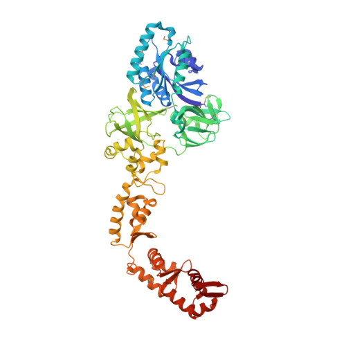
Selenocysteine (Sec), the 21(st) amino acid in translation, uses its specific tRNA (tRNA(Sec)) to recognize the UGA codon. The Sec-specific elongation factor SelB brings the selenocysteinyl-tRNA(Sec) (Sec-tRNA(Sec)) to the ribosome, dependent on both an in-frame UGA and a Sec-insertion sequence (SECIS) in the mRNA. The bacterial SelB binds mRNA through its C-terminal region, for which crystal structures have been reported. In this study, we determined the crystal structure of the full-length SelB from the bacterium Aquifex aeolicus, in complex with a GTP analog, at 3.2-Å resolution. SelB consists of three EF-Tu-like domains (D1-3), followed by four winged-helix domains (WHD1-4). The spacer region, connecting the N- and C-terminal halves, fixes the position of WHD1 relative to D3. The binding site for the Sec moiety of Sec-tRNA(Sec) is located on the interface between D1 and D2, where a cysteine molecule from the crystallization solution is coordinated by Arg residues, which may mimic Sec binding. The Sec-binding site is smaller and more exposed than the corresponding site of EF-Tu. Complex models of Sec-tRNA(Sec), SECIS RNA, and the 70S ribosome suggest that the unique secondary structure of tRNA(Sec) allows SelB to specifically recognize tRNA(Sec) and characteristically place it at the ribosomal A-site.
- RIKEN Systems and Structural Biology Center, 1-7-22 Suehiro-cho, Tsurumi, Yokohama 230-0045, Japan Department of Biophysics and Biochemistry, Graduate School of Science, The University of Tokyo, 7-3-1 Hongo, Bunkyo-ku, Tokyo 113-0033, Japan.
Organizational Affiliation: