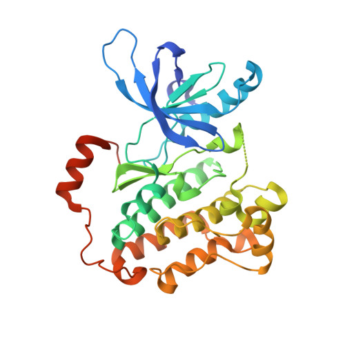
4ZSE

Crystal structure of EGFR 696-1022 T790M/V948R, crystal form II


Experimental Data Snapshot

  • Method: X-RAY DIFFRACTION
  • Resolution: 1.97 Å
  • R-Value Free: 0.213 
  • R-Value Work: 0.190 
  • R-Value Observed: 0.191 

wwPDB Validation   3D Report Full Report


Ligand Structure Quality Assessment 


This is version 1.0 of the entry. See complete history


Literature

Ibrutinib Selectively and Irreversibly Targets EGFR-mutant non-Small Cell Lung Cancer Cells

Wu, H.Wang, A.Zhang, W.Wang, B.Wang, B.Yan, X.E.Chen, C.Hu, C.Ye, Z.Zhao, Z.Wang, L.Li, X.Yu, K.Liu, J.Wu, J.Wang, J.Wang, C.Weisberg, E.L.Liu, J.Gray, N.S.Chen, L.Yun, C.H.Liu, Q.

To be published.

Macromolecules
Find similar proteins by:  (by identity cutoff)  |  3D Structure
Entity ID: 1
MoleculeChainsSequence LengthOrganismDetailsImage
Epidermal growth factor receptor
A, B, C, D
331Homo sapiensMutation(s): 2 
Gene Names: EGFRERBBERBB1HER1
EC: 2.7.10.1
UniProt & NIH Common Fund Data Resources
Find proteins for P00533 (Homo sapiens)
Explore P00533 
Go to UniProtKB:  P00533
PHAROS:  P00533
Entity Groups  
Sequence Clusters30% Identity50% Identity70% Identity90% Identity95% Identity100% Identity
UniProt GroupP00533
Protein Feature View
Expand
  • Reference Sequence
Experimental Data & Validation

Experimental Data

  • Method: X-RAY DIFFRACTION
  • Resolution: 1.97 Å
  • R-Value Free: 0.213 
  • R-Value Work: 0.190 
  • R-Value Observed: 0.191 
  • Space Group: P 1 21 1
Unit Cell:
Length ( Å )Angle ( ˚ )
a = 71.82α = 90
b = 102.317β = 101.24
c = 86.752γ = 90
Software Package:
Software NamePurpose
PHENIXrefinement
PHASERphasing
HKL-3000data processing
Cootmodel building

Structure Validation

View Full Validation Report


Ligand Structure Quality Assessment 


Entry History & Funding Information

Deposition Data


Funding OrganizationLocationGrant Number
China--

Revision History  (Full details and data files)

  • Version 1.0: 2016-06-15
    Type: Initial release