4ZI3 | pdb_00004zi3

BART-like domain of BARTL1/CCDC104 aa1-133 in complex with Arl3FL bound to GppNHp in P1 21 1


Experimental Data Snapshot

  • Method: X-RAY DIFFRACTION
  • Resolution: 2.00 Å
  • R-Value Free: 
    0.243 (Depositor), 0.240 (DCC) 
  • R-Value Work: 
    0.189 (Depositor), 0.190 (DCC) 
  • R-Value Observed: 
    0.192 (Depositor) 

Starting Model: experimental
View more details

wwPDB Validation   3D Report Full Report


Ligand Structure Quality Assessment 

Created with Raphaël 2.3.0Worse 01 BetterLigand structure goodness of fit to experimental dataBest fitted GNPClick on this verticalbar to view details

This is version 1.1 of the entry. See complete history


Literature

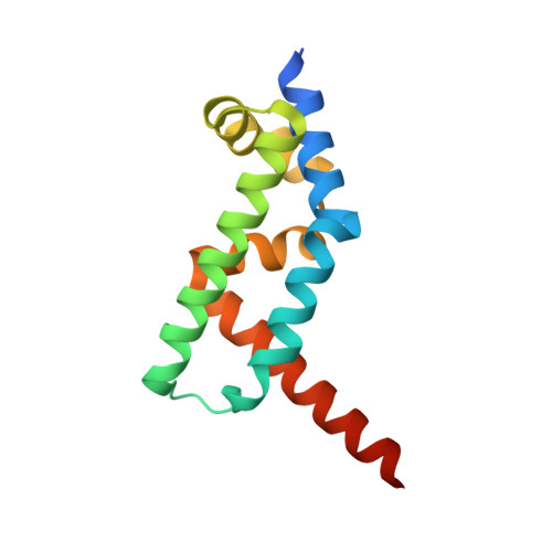
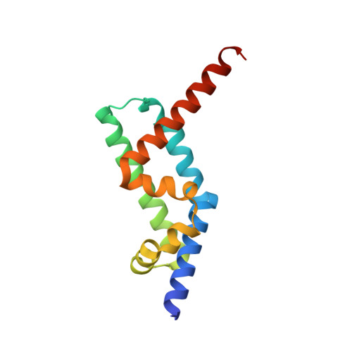
The Interaction of CCDC104/BARTL1 with Arl3 and Implications for Ciliary Function.

Lokaj, M.Kosling, S.K.Koerner, C.Lange, S.M.van Beersum, S.E.van Reeuwijk, J.Roepman, R.Horn, N.Ueffing, M.Boldt, K.Wittinghofer, A.

(2015) Structure 23: 2122-2132

  • DOI: https://doi.org/10.1016/j.str.2015.08.016
  • Primary Citation of Related Structures:  
    4ZI2, 4ZI3

  • PubMed Abstract: 

    Cilia are small antenna-like cellular protrusions critical for many developmental signaling pathways. The ciliary protein Arl3 has been shown to act as a specific release factor for myristoylated and farnesylated ciliary cargo molecules by binding to the effectors Unc119 and PDE6δ. Here we describe a newly identified Arl3 binding partner, CCDC104/CFAP36. Biochemical and structural analyses reveal that the protein contains a BART-like domain and is called BARTL1. It recognizes an LLxILxxL motif at the N-terminal amphipathic helix of Arl3, which is crucial for the interaction with the BART-like domain but also for the ciliary localization of Arl3 itself. These results seem to suggest a ciliary role of BARTL1, and possibly link it to the Arl3 transport network. We thus speculate on a regulatory mechanism whereby BARTL1 aids the presentation of active Arl3 to its GTPase-activating protein RP2 or hinders Arl3 membrane binding in the area of the transition zone.


  • Organizational Affiliation

    Max-Planck-Institute of Molecular Physiology, Emeritus Group, Otto-Hahn-Straße 15, 44227 Dortmund, Germany.


Macromolecules
Find similar proteins by:  (by identity cutoff)  |  3D Structure
Entity ID: 1
MoleculeChains Sequence LengthOrganismDetailsImage
ADP-ribosylation factor-like protein 3
A, B
190Mus musculusMutation(s): 0 
Gene Names: Arl3
UniProt
Find proteins for Q9WUL7 (Mus musculus)
Explore Q9WUL7 
Go to UniProtKB:  Q9WUL7
Entity Groups  
Sequence Clusters30% Identity50% Identity70% Identity90% Identity95% Identity100% Identity
UniProt GroupQ9WUL7
Sequence Annotations
Expand
  • Reference Sequence
Find similar proteins by:  (by identity cutoff)  |  3D Structure
Entity ID: 2
MoleculeChains Sequence LengthOrganismDetailsImage
Cilia- and flagella-associated protein 36135Mus musculusMutation(s): 0 
Gene Names: Cfap36Ccdc104
UniProt & NIH Common Fund Data Resources
Find proteins for Q8C6E0 (Mus musculus)
Explore Q8C6E0 
Go to UniProtKB:  Q8C6E0
IMPC:  MGI:1913994
Entity Groups  
Sequence Clusters30% Identity50% Identity70% Identity90% Identity95% Identity100% Identity
UniProt GroupQ8C6E0
Sequence Annotations
Expand
  • Reference Sequence
Find similar proteins by:  (by identity cutoff)  |  3D Structure
Entity ID: 3
MoleculeChains Sequence LengthOrganismDetailsImage
Cilia- and flagella-associated protein 36133Mus musculusMutation(s): 0 
Gene Names: Cfap36Ccdc104
UniProt & NIH Common Fund Data Resources
Find proteins for Q8C6E0 (Mus musculus)
Explore Q8C6E0 
Go to UniProtKB:  Q8C6E0
IMPC:  MGI:1913994
Entity Groups  
Sequence Clusters30% Identity50% Identity70% Identity90% Identity95% Identity100% Identity
UniProt GroupQ8C6E0
Sequence Annotations
Expand
  • Reference Sequence
Experimental Data & Validation

Experimental Data

  • Method: X-RAY DIFFRACTION
  • Resolution: 2.00 Å
  • R-Value Free:  0.243 (Depositor), 0.240 (DCC) 
  • R-Value Work:  0.189 (Depositor), 0.190 (DCC) 
  • R-Value Observed: 0.192 (Depositor) 
Space Group: P 1 21 1
Unit Cell:
Length ( Å )Angle ( ˚ )
a = 51.55α = 90
b = 67.72β = 102.65
c = 98.47γ = 90
Software Package:
Software NamePurpose
PHENIXrefinement
XDSdata reduction
XSCALEdata scaling
PHASERphasing

Structure Validation

View Full Validation Report


Ligand Structure Quality Assessment 

Created with Raphaël 2.3.0Worse 01 BetterLigand structure goodness of fit to experimental dataBest fitted GNPClick on this verticalbar to view details

Entry History & Funding Information

Deposition Data


Funding OrganizationLocationGrant Number
European Research Council (ERC) Advanced GrantGermanyARCID; No. 268782

Revision History  (Full details and data files)

  • Version 1.0: 2016-03-23
    Type: Initial release
  • Version 1.1: 2024-01-10
    Changes: Data collection, Database references, Derived calculations, Refinement description