4YIS

Crystal Structure of LAGLIDADG Meganuclease I-CpaMI Bound to Uncleaved DNA


Experimental Data Snapshot

  • Method: X-RAY DIFFRACTION
  • Resolution: 2.89 Å
  • R-Value Free: 0.274 
  • R-Value Work: 0.217 
  • R-Value Observed: 0.220 

wwPDB Validation   3D Report Full Report


This is version 1.6 of the entry. See complete history


Literature

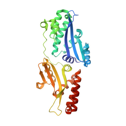
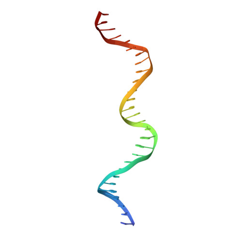
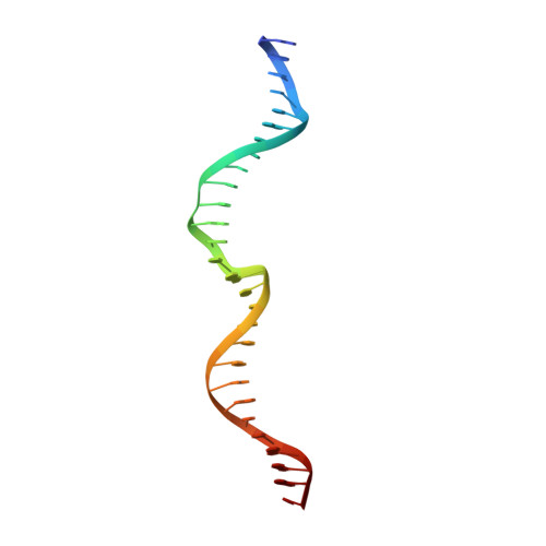
Indirect DNA Sequence Recognition and Its Impact on Nuclease Cleavage Activity.

Lambert, A.R.Hallinan, J.P.Shen, B.W.Chik, J.K.Bolduc, J.M.Kulshina, N.Robins, L.I.Kaiser, B.K.Jarjour, J.Havens, K.Scharenberg, A.M.Stoddard, B.L.

(2016) Structure 24: 862-873

  • DOI: https://doi.org/10.1016/j.str.2016.03.024
  • Primary Citation of Related Structures:  
    4YHX, 4YIS, 4YIT, 4Z1Z, 4Z20, 5ESP

  • PubMed Abstract: 

    LAGLIDADG meganucleases are DNA cleaving enzymes used for genome engineering. While their cleavage specificity can be altered using several protein engineering and selection strategies, their overall targetability is limited by highly specific indirect recognition of the central four base pairs within their recognition sites. In order to examine the physical basis of indirect sequence recognition and to expand the number of such nucleases available for genome engineering, we have determined the target sites, DNA-bound structures, and central four cleavage fidelities of nine related enzymes. Subsequent crystallographic analyses of a meganuclease bound to two noncleavable target sites, each containing a single inactivating base pair substitution at its center, indicates that a localized slip of the mutated base pair causes a small change in the DNA backbone conformation that results in a loss of metal occupancy at one binding site, eliminating cleavage activity.


  • Organizational Affiliation

    Division of Basic Sciences, Fred Hutchinson Cancer Research Center, 1100 Fairview Avenue North, A3-025, Seattle, WA 98109, USA.


Macromolecules

Find similar proteins by:  (by identity cutoff)  |  3D Structure
Entity ID: 1
MoleculeChains Sequence LengthOrganismDetailsImage
Meganuclease I-CpaMI
A, B
295Cryphonectria parasiticaMutation(s): 0 
Gene Names: mtSrRNA I4
UniProt
Find proteins for O20960 (Cryphonectria parasitica)
Explore O20960 
Go to UniProtKB:  O20960
Entity Groups  
Sequence Clusters30% Identity50% Identity70% Identity90% Identity95% Identity100% Identity
UniProt GroupO20960
Sequence Annotations
Expand
  • Reference Sequence
Find similar nucleic acids by:  (by identity cutoff)  |  3D Structure
Entity ID: 2
MoleculeChains LengthOrganismImage
DNA (28-MER)C [auth E],
E [auth C]
28synthetic construct
Sequence Annotations
Expand
  • Reference Sequence
Find similar nucleic acids by:  (by identity cutoff)  |  3D Structure
Entity ID: 3
MoleculeChains LengthOrganismImage
DNA (28-MER)D [auth F],
F [auth D]
28synthetic construct
Sequence Annotations
Expand
  • Reference Sequence
Small Molecules
Ligands 2 Unique
IDChains Name / Formula / InChI Key2D Diagram3D Interactions
EDO
Query on EDO

Download Ideal Coordinates CCD File 
I [auth A],
M [auth B],
N [auth E],
O [auth F],
P [auth D]
1,2-ETHANEDIOL
C2 H6 O2
LYCAIKOWRPUZTN-UHFFFAOYSA-N
CA
Query on CA

Download Ideal Coordinates CCD File 
G [auth A],
H [auth A],
J [auth B],
K [auth B],
L [auth B]
CALCIUM ION
Ca
BHPQYMZQTOCNFJ-UHFFFAOYSA-N
Experimental Data & Validation

Experimental Data

  • Method: X-RAY DIFFRACTION
  • Resolution: 2.89 Å
  • R-Value Free: 0.274 
  • R-Value Work: 0.217 
  • R-Value Observed: 0.220 
  • Space Group: P 21 21 21
Unit Cell:
Length ( Å )Angle ( ˚ )
a = 75.869α = 90
b = 91.799β = 90
c = 139.636γ = 90
Software Package:
Software NamePurpose
HKL-2000data scaling
REFMACrefinement
PDB_EXTRACTdata extraction
Cootmodel building
PHASERphasing

Structure Validation

View Full Validation Report


Entry History & Funding Information

Deposition Data


Funding OrganizationLocationGrant Number
National Institutes of Health/National Institute of General Medical Sciences (NIH/NIGMS)United StatesR01 GM105691

Revision History  (Full details and data files)

  • Version 1.0: 2016-03-23
    Type: Initial release
  • Version 1.1: 2016-03-30
    Changes: Database references
  • Version 1.2: 2016-05-18
    Changes: Database references
  • Version 1.3: 2016-06-15
    Changes: Database references
  • Version 1.4: 2017-09-20
    Changes: Author supporting evidence, Database references, Derived calculations
  • Version 1.5: 2019-12-25
    Changes: Author supporting evidence
  • Version 1.6: 2023-09-27
    Changes: Data collection, Database references, Derived calculations, Refinement description