4XIB

Crystal structure of the MZM-REP domains of Mind bomb 1 in complex with fly Delta N-box peptide


Experimental Data Snapshot

  • Method: X-RAY DIFFRACTION
  • Resolution: 2.15 Å
  • R-Value Free: 0.244 
  • R-Value Work: 0.209 
  • R-Value Observed: 0.212 

wwPDB Validation   3D Report Full Report


This is version 1.5 of the entry. See complete history


Literature

A tail of two sites: a bipartite mechanism for recognition of notch ligands by mind bomb e3 ligases.

McMillan, B.J.Schnute, B.Ohlenhard, N.Zimmerman, B.Miles, L.Beglova, N.Klein, T.Blacklow, S.C.

(2015) Mol Cell 57: 912-924

  • DOI: https://doi.org/10.1016/j.molcel.2015.01.019
  • Primary Citation of Related Structures:  
    4TSE, 4XI6, 4XI7, 4XIB

  • PubMed Abstract: 

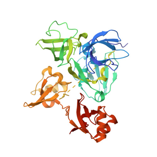
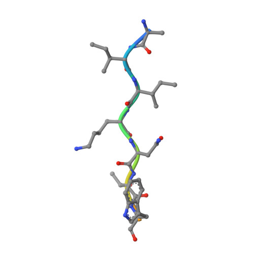
    Mind bomb (Mib) proteins are large, multi-domain E3 ligases that promote ubiquitination of the cytoplasmic tails of Notch ligands. This ubiquitination step marks the ligand proteins for epsin-dependent endocytosis, which is critical for in vivo Notch receptor activation. We present here crystal structures of the substrate recognition domains of Mib1, both in isolation and in complex with peptides derived from Notch ligands. The structures, in combination with biochemical, cellular, and in vivo assays, show that Mib1 contains two independent substrate recognition domains that engage two distinct epitopes from the cytoplasmic tail of the ligand Jagged1, one in the intracellular membrane proximal region and the other near the C terminus. Together, these studies provide insights into the mechanism of ubiquitin transfer by Mind bomb E3 ligases, illuminate a key event in ligand-induced activation of Notch receptors, and identify a potential target for therapeutic modulation of Notch signal transduction in disease.


  • Organizational Affiliation

    Harvard Medical School, Boston, MA 02115, USA.


Macromolecules
Find similar proteins by:  (by identity cutoff)  |  3D Structure
Entity ID: 1
MoleculeChains Sequence LengthOrganismDetailsImage
E3 ubiquitin-protein ligase MIB1380Homo sapiensMutation(s): 0 
Gene Names: MIB1DIP1KIAA1323ZZANK2
EC: 6.3.2
UniProt & NIH Common Fund Data Resources
Find proteins for Q86YT6 (Homo sapiens)
Explore Q86YT6 
Go to UniProtKB:  Q86YT6
PHAROS:  Q86YT6
GTEx:  ENSG00000101752 
Entity Groups  
Sequence Clusters30% Identity50% Identity70% Identity90% Identity95% Identity100% Identity
UniProt GroupQ86YT6
Sequence Annotations
Expand
  • Reference Sequence

Find similar proteins by:  Sequence   |   3D Structure  

Entity ID: 2
MoleculeChains Sequence LengthOrganismDetailsImage
Delta N-box peptideB [auth C]11Drosophila melanogasterMutation(s): 0 
UniProt
Find proteins for P10041 (Drosophila melanogaster)
Explore P10041 
Go to UniProtKB:  P10041
Entity Groups  
Sequence Clusters30% Identity50% Identity70% Identity90% Identity95% Identity100% Identity
UniProt GroupP10041
Sequence Annotations
Expand
  • Reference Sequence
Experimental Data & Validation

Experimental Data

  • Method: X-RAY DIFFRACTION
  • Resolution: 2.15 Å
  • R-Value Free: 0.244 
  • R-Value Work: 0.209 
  • R-Value Observed: 0.212 
  • Space Group: P 41 2 2
Unit Cell:
Length ( Å )Angle ( ˚ )
a = 74.131α = 90
b = 74.131β = 90
c = 170.174γ = 90
Software Package:
Software NamePurpose
PHENIXrefinement
PDB_EXTRACTdata extraction
HKL-2000data scaling
PHENIXphasing
Cootmodel building

Structure Validation

View Full Validation Report


Entry History & Funding Information

Deposition Data


Funding OrganizationLocationGrant Number
National Institutes of Health/National Cancer Institute (NIH/NCI)United StatesP01 CA119070
American Cancer SocietyUnited States120246-PF-11-044-01-DMC
German Research Foundation (DFG)GermanyKL 1028/3-1

Revision History  (Full details and data files)

  • Version 1.0: 2015-03-18
    Type: Initial release
  • Version 1.1: 2015-03-25
    Changes: Database references
  • Version 1.2: 2017-09-20
    Changes: Author supporting evidence, Database references, Derived calculations, Refinement description, Source and taxonomy, Structure summary
  • Version 1.3: 2017-09-27
    Changes: Author supporting evidence
  • Version 1.4: 2019-12-04
    Changes: Author supporting evidence
  • Version 1.5: 2024-02-28
    Changes: Data collection, Database references