4XD7 | pdb_00004xd7

Structure of thermophilic F1-ATPase inhibited by epsilon subunit


Experimental Data Snapshot

  • Method: X-RAY DIFFRACTION
  • Resolution: 3.90 Å
  • R-Value Free: 
    0.282 (Depositor), 0.280 (DCC) 
  • R-Value Work: 
    0.250 (Depositor), 0.250 (DCC) 
  • R-Value Observed: 
    0.251 (Depositor) 

Starting Models: experimental
View more details

wwPDB Validation   3D Report Full Report


Ligand Structure Quality Assessment 

Created with Raphaël 2.3.0Worse 01 BetterLigand structure goodness of fit to experimental dataBest fitted ADPClick on this verticalbar to view details

This is version 1.5 of the entry. See complete history


Literature

Structure of a thermophilic F1 -ATPase inhibited by an epsilon-subunit: deeper insight into the epsilon-inhibition mechanism.

Shirakihara, Y.Shiratori, A.Tanikawa, H.Nakasako, M.Yoshida, M.Suzuki, T.

(2015) FEBS J 282: 2895-2913

  • DOI: https://doi.org/10.1111/febs.13329
  • Primary Citation of Related Structures:  
    4XD7

  • PubMed Abstract: 

    F1-ATPase (F1) is the catalytic sector in F(o)F1-ATP synthase that is responsible for ATP production in living cells. In catalysis, its three catalytic β-subunits undergo nucleotide occupancy-dependent and concerted open-close conformational changes that are accompanied by rotation of the γ-subunit. Bacterial and chloroplast F1 are inhibited by their own ε-subunit. In the ε-inhibited Escherichia coli F1 structure, the ε-subunit stabilizes the overall conformation (half-closed, closed, open) of the β-subunits by inserting its C-terminal helix into the α3β3 cavity. The structure of ε-inhibited thermophilic F1 is similar to that of E. coli F1, showing a similar conformation of the ε-subunit, but the thermophilic ε-subunit stabilizes another unique overall conformation (open, closed, open) of the β-subunits. The ε-C-terminal helix 2 and hook are conserved between the two structures in interactions with target residues and in their positions. Rest of the ε-C-terminal domains are in quite different conformations and positions, and have different modes of interaction with targets. This region is thought to serve ε-inhibition differently. For inhibition, the ε-subunit contacts the second catches of some of the β- and α-subunits, the N- and C-terminal helices, and some of the Rossmann fold segments. Those contacts, as a whole, lead to positioning of those β- and α- second catches in ε-inhibition-specific positions, and prevent rotation of the γ-subunit. Some of the structural features are observed even in IF1 inhibition in mitochondrial F1.


  • Organizational Affiliation

    National Institute of Genetics, Mishima, Japan.


Macromolecules
Find similar proteins by:  (by identity cutoff)  |  3D Structure
Entity ID: 1
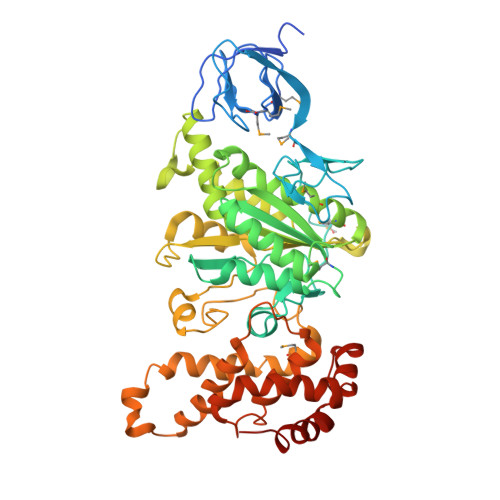
MoleculeChains Sequence LengthOrganismDetailsImage
ATP synthase subunit alpha
A, B, C
502Bacillus sp. PS3Mutation(s): 0 
Gene Names: atpA
EC: 3.6.3.14 (PDB Primary Data), 7.1.2.2 (UniProt)
UniProt
Find proteins for P09219 (Bacillus sp. (strain PS3))
Explore P09219 
Go to UniProtKB:  P09219
Entity Groups  
Sequence Clusters30% Identity50% Identity70% Identity90% Identity95% Identity100% Identity
UniProt GroupP09219
Sequence Annotations
Expand
  • Reference Sequence
Find similar proteins by:  (by identity cutoff)  |  3D Structure
Entity ID: 2
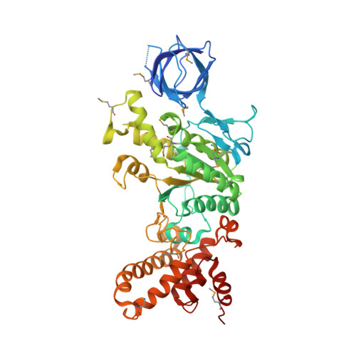
MoleculeChains Sequence LengthOrganismDetailsImage
ATP synthase subunit beta
D, E, F
483Bacillus sp. PS3Mutation(s): 0 
Gene Names: atpD
EC: 3.6.3.14 (PDB Primary Data), 7.1.2.2 (UniProt)
UniProt
Find proteins for P07677 (Bacillus sp. (strain PS3))
Explore P07677 
Go to UniProtKB:  P07677
Entity Groups  
Sequence Clusters30% Identity50% Identity70% Identity90% Identity95% Identity100% Identity
UniProt GroupP07677
Sequence Annotations
Expand
  • Reference Sequence
Find similar proteins by:  (by identity cutoff)  |  3D Structure
Entity ID: 3
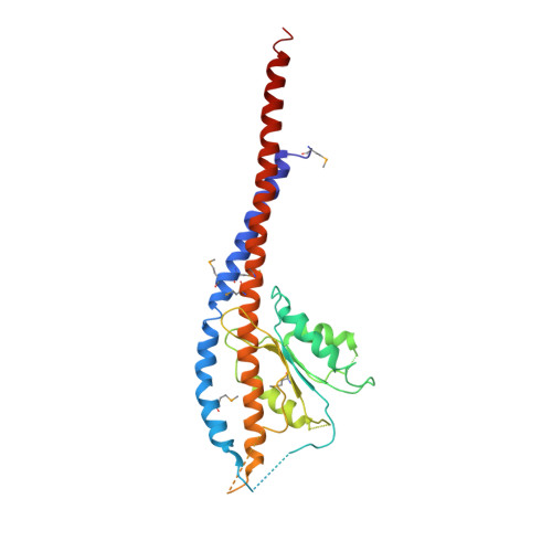
MoleculeChains Sequence LengthOrganismDetailsImage
ATP synthase gamma chain285Bacillus sp. PS3Mutation(s): 0 
Gene Names: atpG
UniProt
Find proteins for P09222 (Bacillus sp. (strain PS3))
Explore P09222 
Go to UniProtKB:  P09222
Entity Groups  
Sequence Clusters30% Identity50% Identity70% Identity90% Identity95% Identity100% Identity
UniProt GroupP09222
Sequence Annotations
Expand
  • Reference Sequence
Find similar proteins by:  (by identity cutoff)  |  3D Structure
Entity ID: 4
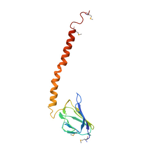
MoleculeChains Sequence LengthOrganismDetailsImage
ATP synthase epsilon chain133Bacillus sp. PS3Mutation(s): 0 
Gene Names: atpC
UniProt
Find proteins for P07678 (Bacillus sp. (strain PS3))
Explore P07678 
Go to UniProtKB:  P07678
Entity Groups  
Sequence Clusters30% Identity50% Identity70% Identity90% Identity95% Identity100% Identity
UniProt GroupP07678
Sequence Annotations
Expand
  • Reference Sequence
Experimental Data & Validation

Experimental Data

  • Method: X-RAY DIFFRACTION
  • Resolution: 3.90 Å
  • R-Value Free:  0.282 (Depositor), 0.280 (DCC) 
  • R-Value Work:  0.250 (Depositor), 0.250 (DCC) 
  • R-Value Observed: 0.251 (Depositor) 
Space Group: I 41 2 2
Unit Cell:
Length ( Å )Angle ( ˚ )
a = 233.395α = 90
b = 233.395β = 90
c = 303.964γ = 90
Software Package:
Software NamePurpose
PHENIXrefinement
MOSFLMdata reduction
SCALAdata scaling
AMoREphasing

Structure Validation

View Full Validation Report


Ligand Structure Quality Assessment 

Created with Raphaël 2.3.0Worse 01 BetterLigand structure goodness of fit to experimental dataBest fitted ADPClick on this verticalbar to view details

Entry History 

Deposition Data

Revision History  (Full details and data files)

  • Version 1.0: 2015-08-26
    Type: Initial release
  • Version 1.1: 2015-10-07
    Changes: Source and taxonomy
  • Version 1.2: 2017-04-19
    Changes: Structure summary
  • Version 1.3: 2023-09-27
    Changes: Advisory, Data collection, Database references, Refinement description
  • Version 1.4: 2023-11-15
    Changes: Data collection
  • Version 1.5: 2024-11-06
    Changes: Structure summary