Structural and Molecular Basis for Resistance to Aminoglycoside Antibiotics by the Adenylyltransferase ANT(2)-Ia.
Cox, G., Stogios, P.J., Savchenko, A., Wright, G.D.(2015) mBio 6
- PubMed: 25564464 Search on PubMedSearch on PubMed Central
- DOI: https://doi.org/10.1128/mBio.02180-14
- Primary Citation Related Structures:
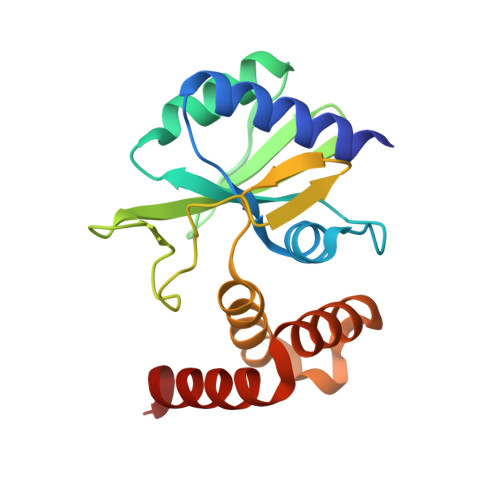
4WQK, 4WQL - PubMed Abstract:
The aminoglycosides are highly effective broad-spectrum antimicrobial agents. However, their efficacy is diminished due to enzyme-mediated covalent modification, which reduces affinity of the drug for the target ribosome. One of the most prevalent aminoglycoside resistance enzymes in Gram-negative pathogens is the adenylyltransferase ANT(2″)-Ia, which confers resistance to gentamicin, tobramycin, and kanamycin. Despite the importance of this enzyme in drug resistance, its structure and molecular mechanism have been elusive. This study describes the structural and mechanistic basis for adenylylation of aminoglycosides by the ANT(2″)-Ia enzyme. ANT(2″)-Ia confers resistance by magnesium-dependent transfer of a nucleoside monophosphate (AMP) to the 2″-hydroxyl of aminoglycoside substrates containing a 2-deoxystreptamine core. The catalyzed reaction follows a direct AMP transfer mechanism from ATP to the substrate antibiotic. Central to catalysis is the coordination of two Mg(2+) ions, positioning of the modifiable substrate ring, and the presence of a catalytic base (Asp86). Comparative structural analysis revealed that ANT(2″)-Ia has a two-domain structure with an N-terminal active-site architecture that is conserved among other antibiotic nucleotidyltransferases, including Lnu(A), LinB, ANT(4')-Ia, ANT(4″)-Ib, and ANT(6)-Ia. There is also similarity between the nucleotidyltransferase fold of ANT(2″)-Ia and DNA polymerase β. This similarity is consistent with evolution from a common ancestor, with the nucleotidyltransferase fold having adapted for activity against chemically distinct molecules. IMPORTANCE : To successfully manage the threat associated with multidrug-resistant infectious diseases, innovative therapeutic strategies need to be developed. One such approach involves the enhancement or potentiation of existing antibiotics against resistant strains of bacteria. The reduction in clinical usefulness of the aminoglycosides is a particular problem among Gram-negative human pathogens, since there are very few therapeutic options for infections caused by these organisms. In order to successfully circumvent or inhibit the activity of aminoglycoside-modifying enzymes, and to thus rejuvenate the activity of the aminoglycoside antibiotics against Gram-negative pathogens, structural and mechanistic information is crucial. This study reveals the structure of a clinically prevalent aminoglycoside resistance enzyme [ANT(2″)-Ia] and depicts the molecular basis underlying modification of antibiotic substrates. Combined, these findings provide the groundwork for the development of broad-spectrum inhibitors against antibiotic nucleotidyltransferases.
- Department of Biochemistry and Biomedical Sciences, Michael G. DeGroote Institute for Infectious Disease Research, McMaster University, Hamilton, Ontario, Canada.
Organizational Affiliation: